Figure 1 CONSORT flow diagram.
Aarti Welling, MPT,1* Ashwin Patil, MD (Radiology),2 Pragati Gunjal, BPT,1 Priyanka Naik, BPT,1 Rani Hubli, BPT1
1Department of Orthopaedic Physiotherapy, KAHER Institute of Physiotherapy, Nehru Nagar, Belagavi 590010, Karnataka, India
2Department of Radiology, J. N. Medical College, KLE Academy of Higher Education and Research, Belagavi, Karnataka, India
Background
Lumbar hyperlordosis is the most prevalent musculoskeletal postural deformity. Maintenance of normal limits of lumbar lordosis is necessary for obtaining an ideal posture. Literature suggests that poor posture results in fascial restriction in which the fascia reorganizes in response to tension. Gross myofascial release (MFR) combined with posterior pelvic tilting exercises proved to be beneficial in improving the lumbar range of motion. Three-dimensional (3D) MFR is a novel approach toward reducing fascial restrictions. However, the literature determining the effects of 3D MFR is still emerging.
Aim
To determine the effect of 3D MFR on a lumbar lordosis angle and lumbar range of motion, in individuals with asymptomatic hyperlordosis.
Method
Participants (n = 30) with hyperlordosis were randomly assigned to either the experimental group receiving 3D MFR (n = 15) or the control group (n = 15) that received sham 3D MFR for six sessions (3 alternate days for 2 weeks). The outcomes were assessed at day 1 and day 6. Lumbar range of motion was assessed using modified-modified Schober’s test and the lumbar lordosis angle was measured using x-ray and flexicurve.
Results
There was significant decrease (p = 0.0001) in the lumbar lordosis angle, increase in the lumbar flexion (p = 0.0001), and decrease in the extension (p = 0.0011) range of motion in the experimental group when compared to the control group.
Conclusion
Lumbar lordosis decreased and the lumbar range of motion increased in the experimental group only with 3D MFR and not with sham 3D MFR. Hence, 3D MFR is an effective method in the correction of lumbar hyperlordosis and improving the lumbar range.
Clinical Trial Registry of India (CTRI) trial number CTRI/2023/03/050340.
KEYWORDS: Lumbar lordosis; fascia; correction; posture
The lumbar spine’s natural curvature, commonly known as lumbar lordosis, plays a pivotal role in maintaining the structural integrity and functional efficiency of the human spine. Deviations from the normal lumbar lordotic curvature can lead to various musculoskeletal issues, impacting an individual’s quality of life.(1,2) Lumbar hyperlordosis is a condition characterized by an excessive inward curvature of the lumbar spine, leading to an exaggerated arching of the lower back. This abnormal posture can result in various biomechanical and structural changes throughout the lumbar region, affecting not only the alignment of the vertebrae but also the surrounding soft tissues, including the muscles and fascia.(3–5) If left untreated, this misalignment places additional stress on the lumbar vertebrae and associated structures, potentially resulting in various issues such as chronic lower back pain, increased risk of disc herniation, muscle imbalances, and gait abnormalities.
Fascia is a connective tissue that surrounds and supports muscles, bones, and organs throughout the body. It is composed of collagen fibers and other extracellular matrix components. Fascia plays a crucial role in maintaining structural integrity, transmitting forces, and providing support to the body.(6) In the context of lumbar hyperlordosis, the increased curvature of the spine and the associated changes in muscle activation and load distribution can create sustained mechanical tension on the lumbar fascia.(7,8) This tension can lead to fascial reorganization, where collagen fibers within the fascia align in response to the altered biomechanical forces. Over time, this reorganization can result in areas of fascial restriction, where the fascia becomes denser and less pliable. These areas of fascial restriction can contribute to reduced mobility and altered muscle function, and potentially exacerbate postural abnormalities associated with lumbar hyperlordosis.(6–8)
Physiotherapy offers diverse treatments for lumbar hyperlordosis, customized to each person’s needs. These encompass tailored exercises, manual therapy, and postural education. Exercises strengthen lumbar-supporting muscles and foster-balanced activation, including core stabilization exercises like pelvic tilts, bridging, and abdominal bracing. Stretching routines target tight hip flexors, lower back muscles, and the chest to counter the exaggerated curve.(9–11) Manual therapy, such as myofascial release (MFR), joint mobilization, and soft-tissue massage, releases tension, eases restrictions, and improves alignment. Postural education highlights ergonomic principles and body mechanics in daily activities to discourage excessive lumbar curvature. The holistic approach hinges on individual biomechanics, aiming to gradually rectify hyperlordosis through exercises, manual techniques, and heightened postural awareness.(12) MFR techniques have emerged as potential therapeutic interventions for addressing musculoskeletal dysfunctions, including those related to spinal curvatures. Manual therapies that focus on the myofascial system, which can be combined with the patient’s active movement, are aimed at improving flexibility and gliding between layers of soft tissues.(6) Stimulation of mechanoreceptors located in the connective tissue may mechanically improve proprioception and reduce the excessive activity of the para-spinal muscles and sympathetic activity of the autonomic nervous system, thus adding significant clinical value to the therapy.(7) Literature suggests gross MFR combined with posterior pelvic tilting exercises proved to be beneficial in improving the lumbar range of motion. Gross MFR techniques in patients with low back pain have proven to decrease pain intensity and increase lumbar flexion and pelvic inclination angle.(12,13)
Traditional MFR involves the application of sustained pressure and stretching to the fascia and muscles to release tension and restrictions. It aims to improve tissue mobility, alleviate pain, and restore normal movement patterns. MFR therapists use various techniques, such as gentle sustained pressure, stretching, and manipulation, to target specific areas of tension and dysfunction in the fascia and soft tissues.(6,14)
Three-dimensional (3D) MFR represents a novel approach in addressing fascial restrictions. 3D MFR is an evolved approach that takes into consideration the 3D nature of the body’s fascial system. It recognizes that fascia exists not only in a linear plane but also in complex multidirectional networks throughout the body. The difference between traditional MFR and 3D MFR is that the traditional form of MFR is typically performed in a two-dimensional manner, focusing on specific planes and directions of tissue tension that involves applying sustained pressure or gentle stretching to specific areas of the body where fascial restrictions or adhesions are believed to be present. 3D MFR is an evolved approach that takes into consideration the 3D nature of the body’s fascial system. It involves a more comprehensive assessment and treatment of fascial restrictions. 3D MFR considers that fascia can have restrictions in multiple planes, including lengthwise, side-to-side, and rotational aspects. Therapists use a variety of techniques and directions to address these multi-planar restrictions that focus on working with the fascial system in three dimensions to address restrictions, imbalances, and dysfunctions.(6) This technique involves performing movements that stretch and release fascial restrictions along multiple planes of movement, creating a more comprehensive and holistic approach to addressing fascial limitations. Through this innovative approach, 3D MFR seeks to promote greater fascial mobility and alleviate restrictions, contributing to improved musculoskeletal function and overall well-being. Lumbar hyperlordosis can create sustained mechanical tension on the lumbar fascia in multiple directions. This tension can lead to fascial reorganization that can alter the biomechanical forces; hence using a 3D approach can have a multidirectional release of the fascia. However, there is dearth in supporting literature proving its effect in reducing lumbar hyperlordosis; hence the study aims to determine the effect of 3D MFR on a lumbar lordosis angle and lumbar range of motion in individuals with asymptomatic hyperlordosis.
This study was conducted as a placebo randomized controlled trial and was registered with the Clinical Trial Registry of India (CTRI) under the trial number CTRI/2023/03/050340. Ethical clearance was obtained from the institutional ethical committee, and written informed consent was collected from all participants. The study procedure adhered to the principles outlined in the Helsinki Declaration of 1975, as revised in 1983. Before the study’s initiation, the purpose and procedures were thoroughly explained to the participants and those willing to take part in the study gave their informed consent.
A total of 55 subjects with hyperlordosis were screened for eligibility, and participants (n = 30) who met the predetermined inclusion criteria were selected. These participants were randomly assigned to either the intervention or control group using a lottery method, maintaining a 1:1 ratio. The enrollment and allocation processes were managed by the investigator responsible for administering the intervention. To ensure comprehensive reporting of the trial, the study followed the CONSORT 2010 statement, including extensions for pilot studies and feasibility trials, as well as the TiDier checklist for maintaining intervention reporting quality (Figure 1).
|
| ||
|
Figure 1 CONSORT flow diagram. | ||
The inclusion criteria were: (i) individuals with asymptomatic lumbar lordosis aged between 18 and 40 years and (ii) a lumbar lordosis angle more than 40°(15) using the flexicurve method. Participants were excluded if (i) they had history of spinal surgeries or spinal fracture, (ii) they were females in the post-partum period, and (iii) they were diagnosed with conditions such as lumbar radiculopathy, lumbar spondylosis, ankylosing spondylitis, and prolapsed intervertebral disc.
The intervention employed in this study followed an individualized approach, and all treatment sessions were conducted in person at the physiotherapy outpatient department (OPD) of a tertiary-care hospital. Notably, all participants successfully completed the intervention protocol, and there were no instances of loss to follow-up throughout the course of the study. The intervention consisted of a total of six sessions over a 2-week period, with each session occurring three times a week on alternate days. The treatment was given in the physiotherapy OPD.
The application of 3D MFR encompassed distinct techniques at three different levels performed bilaterally as outlined below:
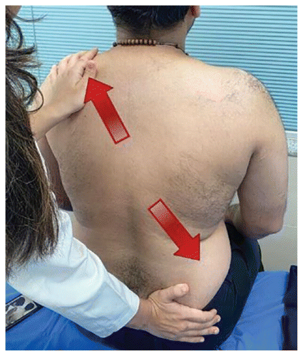
3D stretch (Figure 2): The stretch was performed with the patient sitting on the treatment table and the therapist standing behind the patient. With the hands placed diagonal to each other, stretch was applied laterally and slightly forward at the same time. The stretch was applied bilaterally.
|
| ||
|
Figure 2 Three-dimensional stretch. | ||
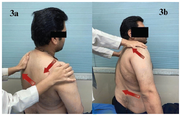
3D stretch with trunk rotation (Figure 3A, 3B): The stretch was performed with the patient sitting on the treatment table and the therapist standing behind the patient. Diagonal stretch downward on one shoulder was applied with one hand and a diagonal stretch up and slightly forward on the other shoulder to initiate the stretch in trunk rotation. In response to the feedback, movement was continued with increased truck rotation and shoulder movement.
|
| ||
|
Figure 3 (A and B) Three-dimensional stretch with trunk rotation. | ||
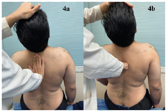
3D stretch with head tilt (Figure 4A, 4B): With the patient in the sitting position, the therapist stands behind the patient. The therapist places the hand on the top of the patient’s head to direct the extension movement of the cervical spine i.e., from anterior to posterior, and simultaneously the therapist applies counter-pressure on the upper thoracic spine with the fist from the posterior to anterior direction after the first stretch. The therapist uses a fist to create a fulcrum and provide counter-pressure. The patient exerts counter-pressure by the back in the backward direction and forward pressure by the head.
|
| ||
|
Figure 4 (A and B) Three-dimensional stretch with head tilt. | ||
The participants received a dosage of 3D MFR involving three sets of release, each lasting for 90 seconds.(6) The treatment session lasted for a duration of 30 minutes.
The control group received sham 3D MFR. While administering sham 3D MFR, the patient’s position and the therapist’s hand placement were same as in the experimental group with a negligible amount of pressure/stretch applied on the skin bilaterally. Identical to the experimental group, the treatment session lasted for a duration of 30 minutes. The intervention consisted of a total of six sessions over a 2-week period, with each session occurring three times a week on alternate days. The treatment was given in the physiotherapy OPD.
All the outcomes were measured two times pre-treatment on day 0 and post-treatment on day 10. The x-ray of the lumbar spine was taken by the technician and the markings of the lordosis angle were performed by a qualified and trained radiologist, whereas the other two outcomes were assessed by a qualified physiotherapist. Both the radiologist and the assessor physiotherapist were blinded to the patient group allocation.
In the context of a research study, the decision to select outcomes related to the degree of lumbar lordosis visualized on x-ray, the magnitude of lumbar curvature measured on the flexicurve, and the lumbar flexion and extension range measured using the modified-modified Schober’s test could be justified based on the comprehensive assessment of lumbar spine health and functionality. These outcomes collectively provide a well-rounded understanding of lumbar spine dynamics and can contribute valuable insights to both clinical practice and research advancements.
Visualizing lumbar lordosis on x-ray allows for a direct assessment of the curvature of the lumbar spine. A lateral radiograph was captured with the patient standing. The patient was instructed to stand comfortably upright while gently resting the fingertips on the same side clavicle and keeping the knees fully extended. The lateral x-ray image encompassed the entire spine from L1 to the sacrum in a single shot. The radiographic procedure was administered by a trained technician. The measurement of the lumbar lordosis angle was conducted by an experienced radiologist. Markings were done on the lateral view of the lumbar spine, and a line was drawn originating from the superior surface of the first lumbar vertebra’s endplate. A second line was then drawn parallel to the upper endplate of the sacral base or the lower endplate of the lowest lumbar segment. Perpendicular lines were subsequently drawn, and the resulting angle at their intersection was measured using a protractor.(16,17)
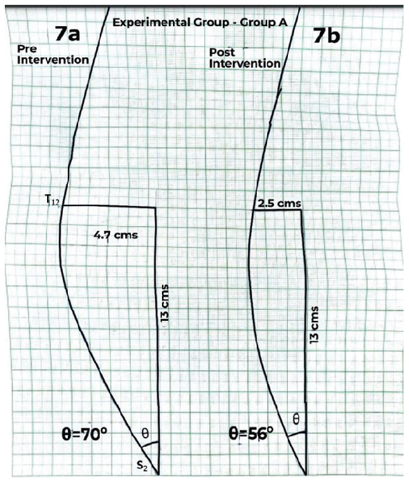
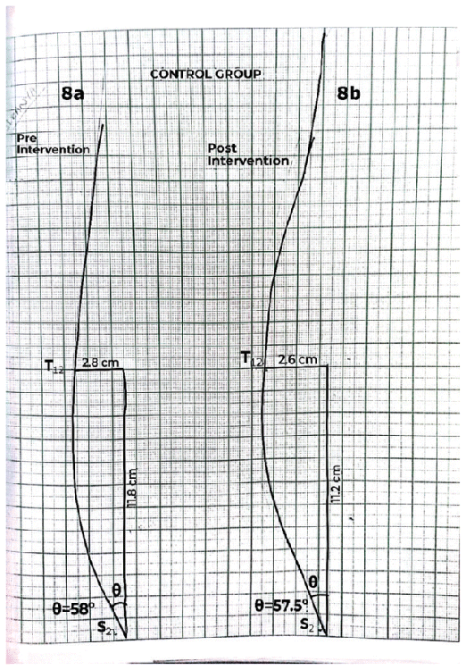
A surveyor’s flexicurve measuring 61 cm in length was used. The flexible ruler was positioned along the spinous processes of the lumbar spinal curve, conformed to the natural curvature of the spine, and then imprinted onto graph paper. This process facilitated the computation of the lordosis index. The utmost breadth and complete extent of the curvature were quantified using the formula θ° = 4 (arc tan [2H/L]); the variable “L” represents the length of a vertical line that connects the T12 and S2 vertebrae, and “H” represents the maximum width or the deepest part of the curvature. This formula is used to quantify the degree of lordotic curvature in the spine, with “H” denoting the depth of the curve and “L” representing the length of the segment along the spine between these two reference vertebrae. The resulting angle (θ°) indicates the extent of the lordotic curve.(18,19)
The patient was positioned upright, with the lower back exposed. Anatomical landmarks, midpoint between posterior superior iliac spines as point A, and a point 15 cm above A as point B were marked. Using a measuring tape, the distance between A and B was noted upright. The patient was asked to bend forward with straight knees. Distance between A and B in this flexed position was measured. The difference between upright and flexed measurements was calculated.(20,21) Similar measurements were taken where the patient was asked to bend backward to measure the extension range of the lumbar spine. The participants were instructed to take off their footwear before the measurements were taken.
Analysis was conducted using IBM SPSS version 23.0. Data normality was assessed via the Kolmogorov–Smirnov test which showed data to be normally distributed; hence a parametric test such as dependent and independent Student’s t-test was used for inferential statistical analysis. The sample size was determined using the formula: n = 2 * S2 * (Zα + Zβ) 2/d2. Here, S represents the standard deviation (S = 3.98), Zα denotes 1.96 at a 5% significance level, Zβ represents 0.842 at 75% power, and d stands for the effect size (d = 3.98, calculated as x1−x2). Based on these values, the calculated sample size was 15 participants per group, totaling 30 participants.
Table 1 represents the demographic characteristics of the participants in the two groups with no significant difference indicating that the participants were matched for their characters.
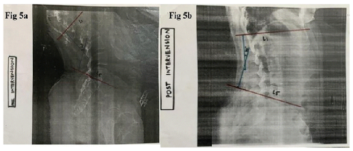
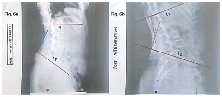
Outcome measure for the within-group analysis according to dependent t-test showed that the % change from pre to post in the experimental group for degree of lumbar lordosis using x-ray was 31.06% (p-value 0.0001), for magnitude of lumbar curvature the % of change was 19.89 (p-value 0.0001), and for the lumbar extension/flexion range the % change was 32.81% and 25.36% (p-value 0.0001 and 0.0011, respectively). The within-group analysis showing % change from pre to post in the sham 3D MFR group for degree of lumbar lordosis using x-ray and lumbar flexion was 0.41% and 2.30% (p-value 0.0824 and 0.334), respectively; there was no significant % of change observed in the magnitude of lumbar curvature and lumbar extension range. The within-group results inferred that the experimental group proved to be superior in terms of decrease in the degree of lumbar lordosis visualized on x-ray (Figure 5A, 5B) when compared to the control group (Figure 6A, 6B) and decrease in the magnitude of lumbar curvature (Figure 7A, 7B) compared to the control group (Figure 8A, 8B) with increase in lumbar flexion and extension range in the experimental group.
|
| ||
|
Figure 5 Pre (A) and post (B) intervention decrease in the degree of lumbar lordosis visualized on x-ray in the experimental group. | ||
|
| ||
|
Figure 6 Pre (A) and post (B) degree of lumbar lordosis visualized on x-ray in the control group. | ||
|
| ||
|
Figure 7 Pre (A) and post (B) decrease in the magnitude of lumbar curvature in the experimental group. | ||
|
| ||
|
Figure 8 Pre (A) and post (B) magnitude of lumbar curvature in the control group. | ||
According to the independent t-test for the between-group analysis, the degree of lumbar lordosis visualized on x-ray, magnitude of lumbar curvature, and lumbar flexion and extension range was seen to have better improvement in group A i.e., experimental group with a p-value of 0.0001 when compared to group B i.e., sham control group (Table 2).
Table 2 Between-Group Analysis Using Independent t-Test for Degree of Lumbar Lordosis, Magnitude of Lumbar Lordosis and Lumbar Flexion/Extension Range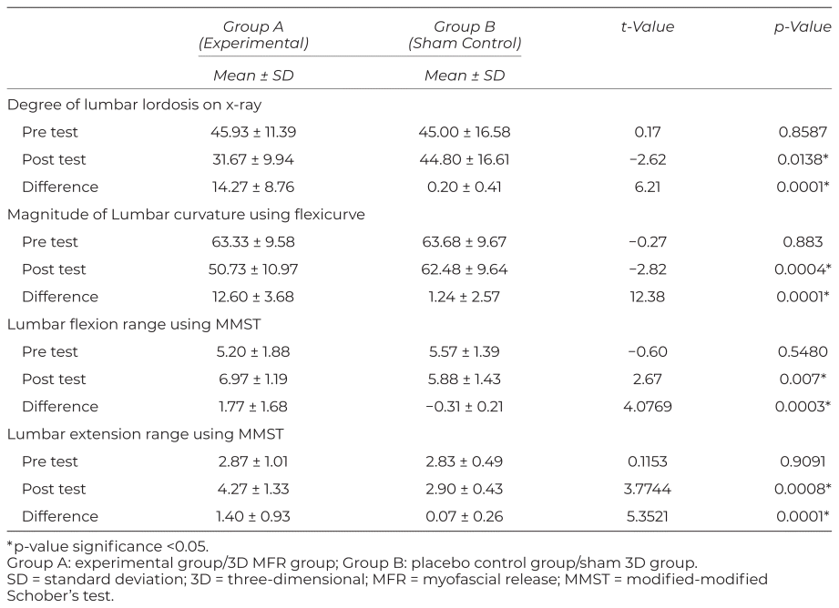
Alternative hypothesis was accepted for all the objective outcome measures used. 3D MFR had a better effect in terms of decrease in the degree of lumbar lordosis visualized on x-ray, decrease in the magnitude of lumbar curvature, and increase in the lumbar flexion and extension range in the experimental group when compared to the sham control group. The present study aimed to determine the effect of 3D MFR on a lumbar lordosis angle and lumbar range of motion, in individuals with asymptomatic hyperlordosis.
The results of the present study showed that there was significant decrease in the degree of lumbar lordosis visualized on x-ray and decrease in the magnitude of lumbar curvature in the experimental group. Firstly, it could be because MFR is a manual therapy technique that aims to release muscle tension and improve fascial mobility.(6) The application of 3D MFR techniques can lead to relaxation of the muscles in the lumbar region, potentially leading to a change in the alignment of the lumbar spine.(12) Secondly, MFR can influence neurological pathways and proprioceptive feedback. Changes in proprioception and neuromuscular control might contribute to alterations in spinal alignment, including the degree of lumbar lordosis.(12)
Existing literature provides evidence suggesting that MFR therapy aims to elongate the fascia, promoting relaxation in soft tissues and joint structures.(12,22) This therapeutic approach contributes to the breakdown of cross-linkages within the soft tissues surrounding the joints. By increasing the space between fibers and enhancing tissue flexibility, MFR facilitates a more comfortable range of motion. Several reviews have affirmed a notable enhancement in lumbar range of motion following the implementation of MFR among patients dealing with chronic low back pain.(22) Interestingly, this study diverges from a previous one suggesting that combining MFR with abdominal exercises or pelvic tilting exercises yields superior outcomes compared to standalone MFR.(23) The divergence could potentially be attributed to the distinctive approach of 3D MFR, which acknowledges the intricate 3D network of the body’s fascial system. 3D MFR acknowledges the fascia’s presence not merely in linear planes but also in intricate, multidirectional networks throughout the body. This approach, in contrast to traditional methods, centers on addressing limitations, imbalances, and dysfunctions by working within the fascial system’s 3D framework. Therapists utilizing 3D MFR incorporate techniques that account for the orientation and interconnectedness of fascial planes in diverse directions.(22,24)
Improvement observed in the flexion and extension range of motion of the lumbar spine in the experimental group could be because, firstly 3D MFR involves working with soft tissues, including fascia, muscles, and connective tissues. Manipulating these structures can lead to changes in tissue length and tension distribution, which in turn might affect the alignment of the lumbar spine. Secondly, there is supporting research indicating that MFR improves flexibility and lengthens myofascial tissue.(22–24) Pollack’s study highlights the significance of fluid dynamics in fascial bodywork. The study introduces the concept of “bound water,” characterized by high viscoelasticity and a trampoline-like bounce. Collagen, a major component of hydrophilic tissue, contains bound water. Restricted fascia shows decreased bound water due to closer collagen and elastin fibers. Photonic energy, like heat from a therapist’s hand, affects this process.(25) Gracovetsky and Chaudhry et al. defined fascia as having nonlinear elastic properties transformable by external forces into heat.(23,24) Other MFR studies correlate interleukin levels with fascial holds. Interleukin 8, regulating inflammation, responds after 3 minutes of holding, doubling at 5 minutes. Interleukin 3, governing blood cell production, rises after a 4-minute hold.(26,27) Considering these findings, MFR’s role in controlling inflammation, enhancing blood flow, reducing pain, and improving myofascial tissue flexibility becomes evident.
Overall, the decrease in the degree, magnitude of lumbar lordosis, and increase in lumbar flexion extension observed after administering 3D MFR can be attributed to the effects of muscle relaxation, changes in soft-tissue tension, alterations in tissue length, and potential neurophysiological influences. 3D MFR places a stronger emphasis on working with the fascial system in three dimensions, taking into account its intricate network throughout the body.
The study’s robustness lies in its capacity to compare the impact of 3D MFR with a placebo control group, effectively establishing and validating the efficacy of 3D MFR. The diverse range of outcomes employed in the study collectively furnished a holistic comprehension of the particulars governing lumbar spine dynamics.
The study’s limitations warrant consideration, including an imbalance in gender distribution among participants, potentially impacting findings and generalizability. Additionally, the absence of a long-term follow-up prevents assessment of sustained effects over time, making the long-term effectiveness and lingering impacts of the 3D MFR approach uncertain.
To conclude, there was decrease in the degree of lumbar hyperlordosis visualized on x-ray, decrease in magnitude of lumbar curvature, and increase in lumbar flexion and extension range only with 3D MFR and not with sham MFR. Hence, 3D MFR is an effective method of releasing the soft tissues and can be used for correction of hyperlordosis.
We thank the radiology department, tertiary-care hospital, Belagavi, for guiding us to mark the angles on x-ray in the department. We thank the statistician Dr. S. B. Javali (Associate Professor in Statistics, USM-KLE International Medical College, Belagavi) for helping us with the analysis of the data.
The authors declare there are no conflicts of interest.
1. Been E, Kalichman L. Lumbar lordosis. Spine J. 2014;14(1):87–97. https://doi.org/10.1016/j.spinee.2013.07.464
Crossref
2. Briggs AM, Greig AM, Wark JD, Fazzalari NL, Bennell KL. A review of anatomical and mechanical factors affecting vertebral body integrity. Int J Med Sci. 2004;1(3):170–180. https://doi.org/10.7150/ijms.1.170
Crossref
3. Roussouly P, Gollogly S, Berthonnaud E, Dimnet J. Classification of the normal variation in the sagittal alignment of the human lumbar spine and pelvis in the standing position. Spine. 2005;30(3):346–353.
Crossref PubMed
4. Guimond S, Massrieh W. Intricate correlation between body posture, personality trait and incidence of body pain: a cross-referential study report. PLoS One. 2012;7:e37450.
Crossref PubMed PMC
5. Shirazi-Adl A, Parnianpour M. Effect of changes in lordosis on mechanics of the lumbar spine-lumbar curvature in lifting. J Spinal Disord. 1999;12(5):436–447.
Crossref PubMed
6. Manheim CJ. In: The Myofascial Release Manual. Thorofare, New Jersey: Slack Incorporated; 2008:4–6.
7. Willard FH, Vleeming A, Schuenke MD, Danneels L, Schleip R. The thoracolumbar fascia: anatomy, function and clinical considerations. J Anat. 2012;221(6):507–536. https://doi.org/10.1111/j.1469-7580.2012.01511.x
Crossref PubMed PMC
8. Sparrey CJ, Bailey JF, Safaee M, Clark AJ, Lafage V, Schwab F, et al. Etiology of lumbar lordosis and its pathophysiology: a review of the evolution of lumbar lordosis, and the mechanics and biology of lumbar degeneration. Neurosurg Focus. 2014;36(5):1–16. https://doi.org/10.3171/2014.1.focus13551
Crossref
9. Varadharajulu G, Bajaj M. Effect of therapeutic exercise protocol in asymptomatic individuals with hyper-lordosis of lumbar spine – an interventional study. Indian J Physiother Occup Ther. 2021;15(2):13–18. Print-(ISSN 0973-5666) and Electronic–(ISSN 0973-5674).
Crossref
10. Bhadauria EA, Gurudut P. Comparative effectiveness of lumbar stabilization, dynamic strengthening, and Pilates on chronic low back pain: randomized clinical trial. J Exerc Rehabil. 2017;3(4):477–485.
Crossref
11. Kudchadkar GS, Gurudut P, Welling A. Comparative effect of mat pilates and egoscue exercises in asymptomatic individuals with lumbar hyperlordosis: a randomized controlled trial. Indian J Phys Ther Res. 2019;1(2):79–88.
Crossref
12. Arun B, Suganya M, Ashok A. Myofascial release therapy in addition to the posterior pelvic tilting in hyperlordosis individuals. Indones J Health Sci. 2019;3(2):71–77.
13. Balasubramaniam A, Mohangandhi V, Sambandamoorthy ACS. Role of myofascial release therapy on pain and lumbar range of motion in mechanical back pain: an exploratory investigation of desk job workers. Ibnosina J Med Biomed Sci. 2014;6(2):75–80.
Crossref
14. Barnes JF. Myofascial release: the missing link in traditional treatment. In: Davis CM, ed. Complementary Therapies in Rehabilitation: Evidence for in Therapy, Prevention, and Wellness. 3rd ed. Thorofare, New Jersey: Slack Incorporated; 2008:89–112.
15. Lin RM, Jou IM, Yu CY. Lumbar lordosis: normal adults. J Formos Med Assoc. 1992;91(3):329–333.
PubMed
16. Yang Z, Xie F, Zhang J, Liang Z, Wang Z, Hu X, et al. An analysis of radiographic parameters comparison between lumbar spine latericumbent and full-length lateral standing radiographs. Spine J. 2017;17(12):1812–1818.
Crossref PubMed
17. Ruhinda E, Byanyima RK, Mugerwa H. Reliability and validity of subjective assessment of lumbar lordosis in conventional radiography. East Afr Med J. 2014;91(10):326–332.
PubMed
18. Seidi F, Rajabi R, Ebrahimi TI, Tavanai AR, Moussavi SJ. The Iranian flexible ruler reliability and validity in lumbar lordosis measurements. World J Sport Sci. 2009;2(2):95–99.
19. de Oliveira TS, Candotti CT, La Torre M, Pelinson PP, Furlanetto TS, Kutchak FM, et al. Validity and reproducibility of the measurements obtained using the flexicurve instrument to evaluate the angles of thoracic and lumbar curvatures of the spine in the sagittal plane. Rehabil Res Pract. 2012;2012:186156.
PubMed PMC
20. Malik K, Sahay P, Saha S, Das RK. Normative values of modified-modified Schober test in measuring lumbar flexion and extension: a cross-sectional study. Int J Health Sci Res. 2016;6:177–187.
21. Amjad F, Mohseni Bandpei MA, Gilani SA, Arooj A. Reliability of modified-modified Schober’s test for the assessment of lumbar range of motion. J Pak Med Assoc. 2022;72(9):1755–1759.
PubMed
22. Tamartash H, Bahrpeyma F. Evaluation of lumbar myofascial release effects on lumbar flexion angle and pelvic inclination angle in patients with nonspecific low back pain. Int J Ther Massage Bodywork. 2022;15(1):15–22.
PubMed PMC
23. Gracovetsky S. Can fascia’s characteristics be influenced by manual therapy? J Bodyw Mov Ther. 2016;20(4):893–897. https://doi.org/10.1016/j.jbmt.2016.08.011
Crossref PubMed
24. Chaudhry H, Bukiet B, Findley T. Mathematical analysis of applied loads on skeletal muscles during manual therapy. J Am Osteopath Assoc. 2008;108(12):680–688.
PubMed
25. Pollack GH. The Fourth Phase of Water. Seattle, WA, USA; Ebner and Sons Publishers; 2013.
26. Meltzer KR, Cao TV, Schad JF, King H, Stoll ST, Standley PR. In vitro modeling of repetitive motion injury and myofascial release. J Bodyw Mov Ther. 2010;14(2):162–171.
Crossref PubMed PMC
27. Standley PR, Meltzer K. In vitro modeling of repetitive motion strain and manual medicine treatments: potential roles for pro- and anti-inflammatory cytokines. J Bodyw Mov Ther. 2008;12:201–203.
Crossref PubMed PMC
Corresponding author: Aarti Welling, MPT, Assistant Professor, Department of Orthopaedic Physiotherapy, KLE Academy of Higher Education and Research Institute of Physiotherapy, Nehru Nagar, Belagavi-590 010, Karnataka, India, E-mail: aartiwell88@gmail.com, Tel: +91-9448814569
COPYRIGHT
Published under the CreativeCommons Attribution-NonCommercial-NoDerivs 3.0 License.
International Journal of Therapeutic Massage and Bodywork, Volume 17, Number 2, June 2024