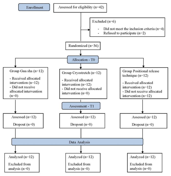
Figure 1 CONSORT flow diagram.
Aditi Jadhav, MPT, Peeyoosha Gurudut, MPT, PhD (Candidate)
Department of Orthopaedic Physiotherapy, KAHER’s Institute of Physiotherapy, KLE Academy of Higher Education & Research, Karnataka, India.Background
Plantar fasciitis (PF) can be treated effectively with manual techniques like cryostretch (CS) and the positional release technique (PRT). Although Gua Sha (GS) has been suggested in the literature for PF, its efficacy has not been studied in the research.
Objective
To determine and compare the effectiveness of GS, CS, and PRT in subjects with PF in terms of pain intensity, pain pressure threshold, and foot function.
Methods/Design
Thirty-six patients with PF (n=36) were randomly allocated to three study groups (12 in each group)—group GS, group CS, and group PRT, respectively.
Settings
A randomized clinical trial was conducted at physiotherapy OPD in a tertiary health center.
Participants
Subjects of all genders with plantar fasciitis of the age group 20–60 years. Thirty-six subjects with plantar fasciitis out of whom 12 were males and 24 females. There were no dropouts in this study.
Intervention
The interventions included the Gua Sha technique (1 session), the cryostretch technique with a frozen tennis ball (3 sessions), and the positional release technique (7 sessions), along with common exercises for all three groups.
Outcome Measures
Pain intensity, foot functions, and pain pressure threshold were assessed using the Numerical Pain Rating Scale, Foot Function Index, and pressure algometer, respectively, on day 1 (pre-intervention) and day 7 (post-intervention).
Results
Between-group analyses showed that group GS was more effective than CS and PRT for pain (p=.0001), group CS was more effective than GS and PRT for foot function (p=.0001) whereas group PRT was more effective than GS and CS for pain pressure threshold (p=.0001).
Conclusion
Although all three groups showed improvement, Gua Sha was superior in terms of reducing pain, cryostretch for improving foot functions, and PRT for reducing tenderness. The interventions used in this study are cost-effective and have proved to be simple and safe techniques.
KEYWORDS: plantar fasciitis, cold therapy, stretching, manual therapy, alternative therapies, soft tissue, manipulation
Heel pain is one of the common foot disorders in which there is extreme discomfort weight-bearing on the heel because of the inflammation of the thick fascia at the sole of the foot. It may be due to either inflammatory or mechanical causes.(1) Plantar fasciitis (PF) is a serious public health issue since it is the common source of heel pain in outpatient department settings. Heel pain affects 10% of the population at some point in their lives, where 83 percent of these patients are active adults who fall under the age group of 25 to 65 years.(2)
The plantar fascia when released has been suggested to be of benefit to patients with symptoms of PF. Various other physiotherapy treatment interventions have been recommended in the past which include rest, taping, cryotherapy, orthotic modifications, silicon heel cups, myofascial release,(3,4) manual stretching,(5) and advice for footwear modifications. Manual therapy is administered in the treatment of PF comprising techniques such as soft tissue mobilizations, deep massage of the tissue, myofascial release, and the positional release technique (PRT).
Gua Sha is a Chinese method of instrument-assisted unidirectional “press-stroking” of a painful area that intentionally creates transitory therapeutic petechiae. These therapeutic petechiae are a result of the eruption of blood into the subcutis and fade within two to five days.(6) The effects of the Gua Sha technique on musculoskeletal conditions like chronic neck pain,(7) low back pain,(8) cervical spondylosis,(9) and lumbar disc herniation(10) have shown better response to pain and functions. A systematic review using Gua Sha to treat musculoskeletal pain concluded insufficient evidence about its effectiveness.(11) To the best of our knowledge and a literature search conducted, there are no studies done to evaluate the effects of Gua Sha on PF subjects.
Cryostretch is a treatment method that includes a combination of cryotherapy and stretching which is given for releasing soft tissues.(12) Myofascial release (MFR) with the help of a tennis ball to the plantar aspect of the foot is widely used to increase flexibility and range of motion further along the posterior muscles.(13) One form of cryostretch technique is given using a frozen tennis ball which helps to release the spasm and increase the muscle/fascia length. The review of literature exhibits lacuna in evidence of the application of the cryostretch technique to confirm its effectiveness in plantar fasciitis.
Positional release technique (PRT), formerly known as strain-counterstrain, is an osteopathic manual therapy technique that aims to improve muscle flexibility by keeping the muscle in a shortened position to promote relaxation of muscle in contrast to placing the muscle in a lengthened or stretched position. PRT as defined by Wynn et al. is an indirect myofascial technique that aims at the neurologic component of the neuro-vascular myofascial somatic dysfunction. PRT is an indirect approach with respect to tissue resistance that includes the use of positioning of the body, utilization of tender points to find the problem and monitoring the therapeutic intervention.(15) In previous studies, PRT has shown a significant decrease in pain and improvement in functional ability in PF patients but has not been compared with the other two treatment approaches.(16)
Further, all three manual therapy interventions work on the basis of different theories/principles. Hence, this study hypothesized that the three techniques, viz. Gua Sha, cryostretch (using a frozen tennis ball), and positional release techniques, will not be equally effective in the management of PF.
The study was approved by the Institutional Research and Ethical Committee with approval number KIPT/SI No.709/07.08.2020. The trial is prospectively registered under the clinical trial registry of India with trial number CTRI/2020/10/028591. Written consent from the subjects was acquired prior to the commencement of the study.
Thirty-six subjects with plantar fasciitis were randomly allocated to one of the three treatment groups with 12 subjects (12 × 3) in each group. The sample size was calculated using the formula where the alpha value was considered as 1.96 at a 5% significance level, the beta value was 1.2816 at 90% power, the standard deviation was 3, and the effect size was set at 4, referring to the previous studies.(11,13) Allocation to the groups was done using the lottery method where participants picked the chits with the group names written on them. The three treatment groups were Gua Sha, Cryostretch, and PRT groups. The inclusion criteria of the study were subjects clinically diagnosed with any one of the following criteria for plantar fasciitis: the painful first step in the morning, calcaneal soreness on palpation, pain on palpation along the proximal plantar fascia,(17) age between 20 and 60 years, and subjects willing to participate in our study. The participants were excluded if they had a recent history of fracture or surgery in and around the ankle joint; congenital deformity of the foot; open wounds, infections; malignancy; sensory impairment, or skin hypersensitivity. All the subjects completed the study intervention with no loss to follow-up (Figure 1).
|
| ||
|
Figure 1 CONSORT flow diagram. | ||
The interventions were carried out by a qualified physiotherapist who had additional certification in the application of instrument-assisted soft tissue manipulation techniques with jade stone, as well as in positional release therapy.
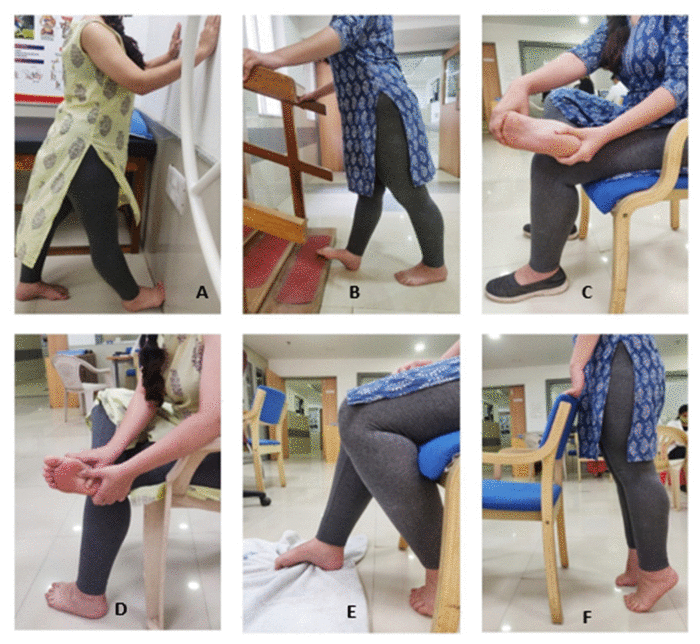
The common intervention comprised of exercises prescribed for targeting the ankle and foot: standing calf stretch (15–30 sec, 3 sets), curb/stair stretch (15 sec, 3 sets), seated plantar fascia stretch (15 sec, 3 sets), cross-friction massage above the plantar fascia (3–5 min), toe curls (10–20 reps), and heel raises (15 reps, 5-sec holds, 2 sets). The materials required for the following exercises were: a chair, stairs/curb, and a towel. The standing calf stretch and curb/stairs stretch focused on the gastrocnemius and soleus muscles stretching. The plantar fascia stretches and cross-friction massage above the plantar fascia are beneficial for walking to help stretch and warm up the fascia before the first steps of the day. The exercises were performed by the patients sitting on a chair. The toe curls and heel raises are strengthening exercises focused on the foot’s intrinsic muscles. Both of these exercises were performed in a standing position with the subject’s hands resting on a chair. The toe curls were performed with the help of a towel, and for the heel raises, the subjects were asked to raise their heels off the floor while keeping their knees straight. The subjects performed these exercises twice a day, once in presence of the therapist in the OPD and the second time at home (Figure 2).(18)
|
| ||
|
Figure 2 Common exercises (A-standing calf stretch, B-curb/stair stretch, C-seated plantar fascia stretch, D-cross-friction massage above the plantar fascia, E-toe curls, F-heel raises). | ||
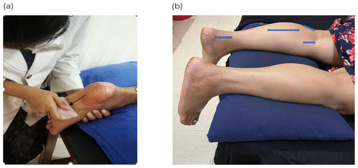
Group GS received the Gua Sha technique along with exercises included in the common intervention. This technique was performed using a jade stone and a skin lubricant to decrease friction. Gua Sha technique was anciently done using a ceramic Chinese soup spoon or a blunt, well-worn coin. Today practitioners commonly use jade or rose quartz or both. However, considering the cost-effectiveness of jade stone which has similar effects as that of rose quartz, in addition to the training involved in the application of jade stone by the therapist involved in the intervention, the jade stone was chosen for the application of Gua Sha. The subjects were treated in a prone lying position where the therapist was standing near the affected foot of the subject. There was sequential press unidirectional stroking along the orientation of the fascia from heel to toe direction, followed by areas including tendon Achilles, calf muscle bulk, and origin of gastrocnemius with 3 min of stroking per area.(19,20) This technique was administered only once, on the first day of the intervention, because the recommended dosage is 1 session per seven days considering the petechial rashes that result due to the technique. The remaining six days were continued only with the exercises (Figure 3).
|
| ||
|
Figure 3 (a) Gua Sha for plantar fascia with jade stone; (b) Gua Sha for tendoachilles, calf and gastrocnemius muscle origins. | ||
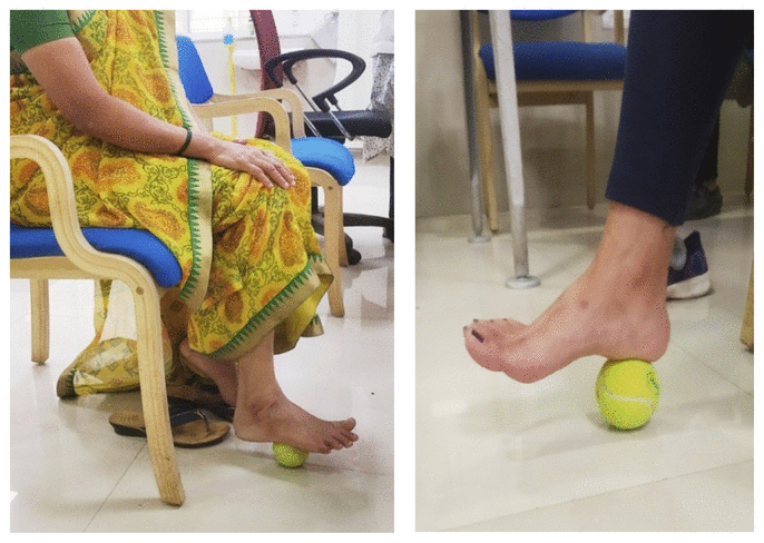
Group CS received the cryostretch technique along with the exercises mentioned above in the section on common intervention. This technique was performed using a frozen tennis ball. The subjects were sitting seated on a chair where one foot was on the frozen tennis ball and the other was flat on the floor. The subjects were then asked to roll the frozen ball under the arch of their foot extending from the heel to the metatarsal heads concentrating on the medial arch for 2 min (30 reps).(13) The subjects were instructed to apply as much pressure as they could, pushing into discomfort but not pain, as greater pressures have shown to have better benefits on flexibility. This technique was administered on alternate days (the 1st, 3rd, and 5th day of the week) along with the exercises (Figure 4).
|
| ||
|
Figure 4 Cryostretch technique. | ||
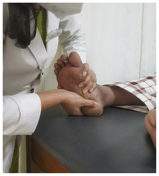
Group PRT received positional release technique along with exercises. The subjects were made to lie in a supine position with the affected limb out of the plinth and then slight mechanical pressure was put on the tender point with one fingertip to determine the tenderness. The foot was then positioned into complete plantar flexion and gently fine-tuned by rotation until there was a reduction in tender point by at least 70%, and this position was held for 90 sec with 3 repetitions.(21) The subjects received this treatment for 7 consecutive sessions over a period of seven days, along with the exercises (Figure 5).
|
| ||
|
Figure 5 Positional release technique. | ||
Outcome measures included in the study were pain intensity using a Numerical Pain Rating Scale (NPRS), foot functions using Foot Function Index (FFI), and pain pressure threshold using a pressure algometer. The demographic data of each subject were documented along with the initial assessment of the outcome measures prior to the intervention (baseline) and post-intervention (Day 7). The duration of the intervention was one week.
The NPRS was used to measure pain intensity. The subjects were asked to indicate the numeric value on an 11-point numerical scale (horizontal bar or line format) according to their current pain over the past 24 hours where 0 was considered as “No Pain” and 10 was considered as “worst Pain’ possible. The NPRS is a valid scale to measure pain intensity and the reliability of this scale is 0.90.(22)
Foot Function Index (FFI) is a self-administered index consisting of 23 items divided into three sub-scales: pain subscale, disability subscale, and activity limitation subscale. The subject was asked to score each question on a scale from 0 (no pain or difficulty) to 10 (worst pain imaginable or so difficult it requires help), which best describes their foot functional ability over the previous week. The FFI is a valid and reliable tool for patients with non-traumatic foot or ankle problems. Test/retest reliability of the FFI total and sub-scale scores ranged from 0.87 to 0.69, whereas the internal consistency ranges from 0.96 to 0.73.(23)
The pain pressure threshold was assessed using a pressure algometer (Baseline brand; White Plains, NY) which is a hand-held instrument where the pressure threshold at which the patient feels the pain is calibrated in kg/cm2 with a range of 10 kg and 0.1 kg divisions. The subjects were asked to point to the location of the worst pain on the foot to identify the maximum tender spot (TS). The subject was asked to confirm pain or discomfort at the point. The indicated site was then palpated by the examiners to identify the correct TS which was marked with a marker. The tip of the algometer was kept over the marked area of maximum tenderness, perpendicular to the involved muscle/fascia. The pressure was progressively increased and the readings at which the subject felt pain were recorded.(24)
Statistical analysis for the present study was done using SPSS version 23.0 (IBM SPSS Statistics, Armonk, NY, USA) to verify the results obtained. Data were summarized as mean ± standard deviation for continuous variables, whereas the categorical variables were represented as a percentage. A comparison of the difference between the three groups was done using a one-way analysis of variance (ANOVA) while within-group differences were analyzed with repeated measures ANOVA.
Details on descriptive statistics that include gender, the side affected, age, body mass index (BMI), duration of pain/symptoms, and the baseline characteristics of the subjects are presented in Table 1.
Table 1 Demographic Profile of Three Study Groups (Group GS, Group CS and Group PRT)
Assessment of pain intensity demonstrated a statistically significant reduction in pain on Day 7 post-intervention in all three groups at pre- to post-intervention (p<.001), as well as between the three groups (p<.001). Similarly, the functional disability (p<.001), as well as pain pressure threshold (p<.001), also showed statistically significant improvements on Day 7 post-intervention. In terms of improved Foot Function Index (p<.001) and pain pressure threshold (p<.001), there was a significant difference between the three study groups (Table 2). The percentage changes were 30, 17.28, and 16.28 for NPRS scores, 10, 17.90, and 9.66 for FFI scores, and 15.12, 9.94, and 33.66 for pressure pain threshold for GS, CS, and PRT groups. respectively.
Table 2 Outcome Measure Scores for Gua Sha (GS), Cryostretch (CS), and Positional Release Technique (PRT)a
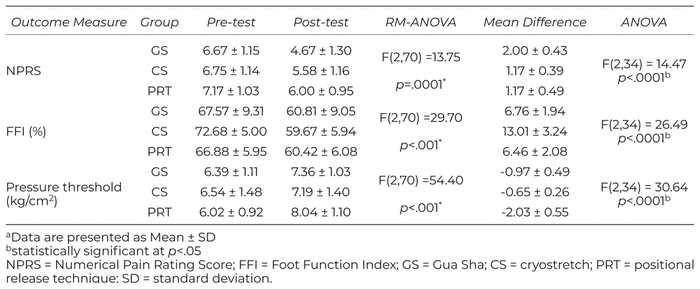
Further, in comparison of the three groups for the mean difference (GS, CS, and PRT), group GS was better than group CS (p<.001) and group PRT (p<.001) for pain intensity outcome. Group CS was demonstrated to be significantly better than group GS (p<.001) and group PRT (p<.001) for Foot Function Index to assess foot disability. Group PRT resulted to be superior to group GS (p<.001) and group CS (p<.001) in terms of pain pressure threshold outcome.
The present study compared the effectiveness Gua Sha, cryostretch, and positional release therapy in subjects with plantar fasciitis in terms of pain intensity, pain threshold, and function. The results of the current study indicated that all three groups (GS, CS, and PRT) showed statistically significant improvement in terms of reducing pain intensity, improving functional ability, and reducing tenderness on Day 7 post-intervention. The results also indicate that group GS was statistically better than CS and PRT in terms of pain; group CS was better than GS and PRT in terms of functional disability; and group PRT was better than GS and CS in terms of pain pressure threshold.
The results of the study showed there was a significant difference in group GS in terms of pain intensity and functional disability on Day 7 post-intervention. GS is known to have beneficial short-term effects on pain intensity; firstly because of the nociceptive activation that plays a role in pain. GS helps in reducing pain through anti-nociceptive effects and also by counter-irritation. Secondly, Gua Sha therapy might have a powerful placebo effect on pain. Thirdly, GS increases local microcirculation, hence decreasing myalgia.(25) GS leads to the circulation of nitric oxide. Nitric oxide also has effects on platelet function, inflammation, and pain perception. Studies have also shown that GS resolves spasms and pain and enhances circulation to the muscles, as well as tissues and organs directly below the area that is treated. Research also states that there is an immediate reduction in stiffness and pain experienced by the patient with increased mobility.(19)
Studies have reported a decrease in pain intensity with the help of GS in the chronic neck pain population, low back pain, musculoskeletal pain, breast engorgement, cervical spondylosis, and fibromyalgia syndrome.(10–13,15) Further, jade stone mobilization reduces muscle spasms and increases blood flow to the treatment area, which has positive implications for the outcome of this study.(19,20) Evidence is lacking regarding the effects of the GS technique on functional disability in plantar fasciitis patients. Since the results of the current study correlate with previous studies, which demonstrate a decrease in functional disability and pain, it can be said that GS is beneficial to improve the functional status of patients with plantar fasciitis.
In the present study, there was a significant improvement in the pain pressure threshold in group GS on day 7 post-intervention. Due to GS therapy, there is a rise in the surface micro-perfusion after the treatment that leads to an eruption of blood in the capillary bed which is associated with an up-regulation of the heme oxygenase-1 (HO-1) gene expression. This up-regulation of HO-1 has anti-nociceptive effects, and anti-inflammatory and immune-regulatory properties which result in an increase in pain pressure thresholds.(26) Since the results of the current study have seen similar effects of an increase in the PPT which supports the previous studies, we can say that GS can be used to increase the PPT in patients with PF.
The common types of stones used for Gua Sha include either rose quartz or jade stone. The selection of the jade stone in the present study was based on its availability in the study location and the training of the therapist who gave the intervention. There are differences in the feel and cost of the two stones. Firstly, rose quartz is more expensive as compared to jade stone. Also, jade is a softer stone whereas rose quartz is harder and cooler. There is evidence of both jade stone and rose quartz being applied for the Gua Sha technique. However, the difference in their outcomes based on the type of stone used has not been studied in the past with comparative trials.
The results of our study showed that there was a significant reduction in group CS in terms of pain intensity, functional disability, and an increase in terms of PPT on Day 7 post-intervention. Cryotherapy was found to be effective in relieving pain by decreasing the inflammation around the plantar fascia. Cryotherapy reduces local hyperthermia, induces vasoconstriction, and lowers microcirculation. The eruption of blood into the surrounding tissues decreases local inflammation, and edema production is also reduced, along with a decrease in motor as well as sensory nerve conduction.(27) Stretching improves blood circulation which brings nutrients to cells and there is the removal of waste products. The rise in the blood flow causes the opening of the connective tissues, which helps alleviate pain.(28)
Previous studies have found that the increase in PPT following stretching with a tennis ball in the ipsilateral and contralateral plantar flexors may be because of the mechanical pressure that rolling tennis balls exert on mechanoreceptors and proprioceptors.(29) Mechanical stress like massage, which removes trigger points from muscle tissue, leads to increased PPT. The application of mechanical pressure applied on trigger points averts unwanted firing of muscle spindles from the trigger point, decreases muscle spasms, and helps to decrease the pain.(30)
In the current study, there was a significant difference in the PRT group in terms of pain intensity and functional disability on Day 7 post-intervention. The factors that helped in pain reduction may be explained due to the reduction in the intra and extrafusal fiber difference and reset of the inappropriate proprioceptive activity. PRT uses static ischemic compression, a position of comfort, and fine-tuning on reflexogenic trigger points to resolve the associated dysfunction.(15)
Positional release therapy is a technique suggested to improve muscle flexibility by keeping the muscle in a shortened position, which inhibits muscle relaxation, rather than keeping the muscle in a lengthened or stretched condition. The utilization of body positioning, tender points to find the problem, and monitoring of the intervention are some of the PRT indirect approaches regarding tissue resistance. The excitability of the myotatic reflex arc might lead to restriction in the movement, which is caused by excessive gamma gain. By keeping the patient’s muscle in the placement of ease for a small period of time, there is a decrease in gamma gain; thereby allowing the excited reflex arc to return back to its original state and increase the range of motion.(16)
The PRT group demonstrated that trigger point sensitivity decreased post-intervention. Our study results agreed with another study where PRT was used in decreasing tender points by increasing pressure pain thresholds of trigger points in the upper trapezius muscle with mechanical neck pain patients.(31) The application of PRT causes a decrease in tissue tenderness by changing nociceptor activity in the soft tissues. The increase in pain pressure threshold is associated with a reduction in the susceptibility of the tissue. Based on previous literature and our current findings, PRT interventions have the ability to relieve tenderness and local pain caused by myofascial trigger points.(32)
However, there were a few limitations to our study such as the lack of a control group, the short duration of the intervention, and no long-term follow-up to assess the continuity of practice that needs to be considered in future studies. Also, there are more chances of a higher number of patient/therapist interactions in the CS and PRT intervention groups than the GS group due to a greater number of treatment sessions in a week which may influence pain perception. A follow-up period can be used to see the long-term effects of all three techniques. More standard outcome measures, like ultrasonography at pre and post-intervention comparison, can be used for a better understanding of the results.
The present study concluded that all three groups—Gua Sha, cryostretch, and positional release techniques—demonstrated significant improvement in terms of reducing pain intensity and foot function score, and increasing pain pressure threshold on Day 7 post-intervention. Further, the Gua Sha intervention was more effective than the cryostretch and positional release technique for pain; the cryostretch technique was more effective for foot function scoring; whereas the positional release technique was more effective for pain pressure threshold. To the best of our knowledge, our study is the first to evaluate the effectiveness of the Gua Sha technique on plantar fasciitis patients. Level II evidence is created by this study where three different manual therapies have been compared. Further, the interventions used in this study are cost-effective and have proved to be simple and safe techniques.
We are thankful to all the subjects for their cooperation. We would also like to thank our statistician, Dr. Manjunath Javali, for helping us with the statistical analysis of this study.
The authors declare there are no conflicts of interest.
1 Borland A, Martin CH, Locke J. Nurses’ understandings of suitable footwear for older people. Int J Healthcare Qual Assur. 2013;26(7):633. Available from: https://www.researchgate.net/publication/257012954_Nurses'_understandings_of_suitable_footwear_for_older_people
2 Scher D, Belmont P, Owens B. The epidemiology of plantar fasciitis. Lower Extremity Rev. 2010;12(1):60
3 Ajimsha MS, Binsu D, Chithra S. Effectiveness of myofascial release in the management of plantar heel pain: a randomized controlled trial. The Foot. 2014;24(2):66–71.


4 Kuhar S, Subhash K, Chitra J. Effectiveness of myofascial release in treatment of plantar fasciitis: A RCT. Indian J Physiother Occup Ther. 2007;1(3):3–9.
5 Orchard J. Plantar fasciitis. BMJ. 2012;345(1): 6603–6603.
6 Lauche R, Wübbeling K, Lüdtke R, Cramer H, Choi KE, Rampp T, et al. Randomized controlled pilot study: pain intensity and pressure pain thresholds in patients with neck and low back pain before and after traditional East Asian” gua sha” therapy. Am J Chinese Med. 2012;40(05):905–917.
7 Braun M, Schwickert M, Nielsen A, Brunnhuber S, Dobos G, Musial F, et al. Effectiveness of traditional Chinese “gua sha” therapy in patients with chronic neck pain: a randomized controlled trial. Pain Med. 2011;12(3):362–369.
8 Puttaswamaiah R, Chandran P. Degenerative plantar fasciitis: a review of current concepts. The Foot. 2007;17(1):3–9.
9 Ma H, Li SZ, Zheng HM: Clinical observation gua sha plus herbal injection with 83 cases of cervical spondylosis. Zhengjiu Lingchuang Zazhi. 2003;19:27–28.
10 Wang ZG, Tao Y, Wu NT. The effect of coin scraping therapy for the treatment of lumbar disc herniation. Zhongguo Zhongyi Gushangke Zazhi. 2004;12:7–10.
11 Lee M, Choi TY, Kim JI, Choi SM. Using Guasha to treat musculoskeletal pain: a systematic review of controlled clinical trials. Chinese Med. 2010;5(1): 1–5
12 Yadav M, Attrey P, Kashyap P. Comparative study between cryostretch and light concentric exercise on delayed onset muscle soreness. Indian J Phys Educ Sports Appl Sci. 2016;44975451.
13 Grieve R, Goodwin F, Alfaki M, Bourton AJ, Jeffries C, Scott H. The immediate effect of bilateral self-myofascial release on the plantar surface of the feet on hamstring and lumbar spine flexibility: a pilot randomised controlled trial. J Bodywk Move Ther. 2015;19(3):544–52.
14 Wynne MM, Burns JM, Eland DC, Conatser RR, Howell JN. Effect of counterstrain on stretch reflexes, Hoffman reflexes, and clinical outcomes in subjects with plantar fasciitis. J Osteopath Med. 2006 Sep 1;106(9):547–556.
15 D’Ambriogo K, Roth G. Text Book of Positional Release Therapy, Assessment and Treatment of Musculoskeletal Dysfunction, 2nd ed. Philadelphia: Mosby; 2002. p.268–282
16 Unver B, Erdem E, Akbas E. Effects of short-foot exercises on foot posture, pain, disability, and plantar pressure in pesplanus. J Sport Rehabil. 2019;29(4):436–440.
17 Hoebeke RE. Diagnosing plantar fasciitis. J Nurs Pract. 2008;4(1):66–67.
18 Roxas M. Plantar fasciitis: diagnosis and therapeutic considerations. Altern Med Rev. 2005;10(2): 83–93.
19 Nielsen A. Gua sha: a Traditional Technique for Modern Practice, 2nd. ed. [e-book]. Amsterdam: Elsevier Health Sciences; 2014.
20 Smith T. Plantar Fasciitis & Gua Sha [video]. Arroyo Grande Health & Wellness [Internet]. 2012. [cited 20 November 2019]. Available from: https://youtu.be/1YH3EbGANi4
21 Am H, Kage Vijay B, Basavaraj C. Comparison of myofascial release and positional release therapy in plantar fasciitis—a clinical trial. Physiother Occup Ther. 2010;4(4):8
22 Krebs EE, Carey TS, Weinberger M. Accuracy of the pain numeric rating scale as a screening test in primary care. J Gen Intern Med. 2007;22(10): 1453–1458.
23 Soo Hoo NF, Samimi DB, Vyas RM, Botzler T. Evaluation of the validity of the Foot Function Index in measuring outcomes in patients with foot and ankle disorders. Foot Ankle Int. 2006;27(1): 38–42.
24 Imamura M, Fischer AA, Imamura ST, Kaziyama HS, Carvalho AE, Salomao O. Treatment of myofascial pain components in plantar fasciitis speeds up recovery: documentation by algometry. J Musculoskel Pain. 1998;6(1):91–110.
25 Nielsen A, Knoblauch NT, Dobos GJ, Michalsen A, Kaptchuk TJ. The effect of Gua Sha treatment on the microcirculation of surface tissue: a pilot study in healthy subjects. Explore. 2007;3(5):456–466.
26 Nascimento C, Branco L. Role of the peripheral hemeoxygenase–carbon monoxide pathway on the nociceptive response of rats to the formalin test: Evidence for a cGMP signaling pathway. Eur J Pharmacol. 2007;556(1–3):55–61.
27 Block, J. Cold and compression in the management of musculoskeletal injuries and orthopedic operative procedures: a narrative review. J Sports Med. 2010;1:105–113.
28 Mohamed, H. Effectiveness of Achilles tendon stretching for the treatment of chronic plantar fasciitis. Egypt Orthopaed J. 2015;50(4):215.
29 Aboodarda SJ, Spence AJ, Button DC. Pain pressure threshold of a muscle tender spot increases following local and non-local rolling massage. BMC Musculoskelet Disord. 2015;16(1):1.
30 Kostopoulos D, Nelson J, Arthur J, Ingber RS, Larkin RW. Reduction of spontaneous electrical activity and pain perception of trigger points in the upper trapezius muscle through trigger point compression and passive stretching. J Muscoskelet Pain. 2008;16(4):266–278.
31 AL-Shawabka SA, Shenouda MM, Balbaa AA. Positional release technique versus manual pressure release on the upper trapezius muscle in patients with myofascial pain dysfunction syndrome. Bull Fac Phys Ther. 2013;18(1):55–63.
32 Weiselfish S. Manual Therapy for the Orthopedic and Neurologic Patient Emphasizing Strain and Counterstrain Technique. Hartford, CT: Regional Physical Therapy (self-published); 1993.
COPYRIGHT
Published under the CreativeCommons Attribution-NonCommercial-NoDerivs 3.0 License.
INTERNATIONAL JOURNAL OF THERAPEUTIC MASSAGE AND BODYWORK, VOLUME 16, NUMBER 1, MARCH 2023