Kathleen (Kate) Zink, MSN, RN, AHN-BC, LMT, [retired]1*, Barbara Chini, MD2, Joyce Cowens, AD, LMT4, Lois Kremer, BSN, RN, [retired]1, Li Lin, MS, BS3
1Division of Child Life & Integrative Care, Cincinnati Children’s Hospital Medical Center, Cincinnati, OH, USA
2Division of Pulmonary Medicine, Cincinnati Children’s Hospital Medical Center, Cincinnati, OH, USA
3Patient Services, Research, Cincinnati Children’s Hospital Medical Center, Cincinnati, OH, USA
4All About Health, Cincinnati, OH, USA
Background
Cystic Fibrosis (CF) is an autosomal recessive disorder of exocrine glands characterized by abnormal production of thick mucus, primarily in bronchi of the lungs. Individuals experience recurrent respiratory infections, increased work of breathing, cough and musculoskeletal changes with pain. Previous research found that massage therapy (MT) decreased pain, muscle tightness, and anxiety in individuals with CF, but did not use valid/reliable measurements of quality of life (QOL).
Purpose
To evaluate the effects of MT on QOL and clinical outcomes in individuals 8 to 21 years old with CF.
Setting
A 622-bed nonprofit pediatric hospital in Ohio in the United States.
Participants
Convenience sample of 24 patients with CF; 12 randomly assigned to treatment and control groups, respectively.
Research Design and Intervention
Prospective two-group controlled pre/post pilot study using deep tissue myofascial trigger point massage over 10 to 12 weeks.
Measurements
Pediatric Quality of Life Inventory (Peds QL 4.0); Cystic Fibrosis Questionnaire-Revised (CFQ-R); numeric rating scales (NRS) for pain, muscle tightness, ease of breathing, relaxation; pulmonary function (PFT); single breath count; thoracic excursion (TE).
Results
All participants were Caucasian; mean age 15.7 (SD = 3.5) years; 16 (66.6%) female. No significant differences were found in terms of age, gender, baseline pain between MT and control groups. At the final visit, compared to the control group, the children in MT group showed statistically significantly reduced muscle tightness (p = .048) with a large effect size (ω2 =0.163) and marginally statistically significantly higher levels of relaxation (p = .052), less pain (p = .076), and improved upper TE (p = .078) and lower TE (p = .056) scores with large and moderate effect sizes (ω2 = 0.156, ω2 = 0.095, ω2 = 0.083, and ω2 = 0.073). No statistically significant differences in children’s and caregivers’ QOL scores between the two groups were found.
Conclusions
Massage therapy was found to significantly reduce muscle tightness, marginally significantly help pain, relaxation, and thoracic excursion in participants with CF.
KEYWORDS: cystic fibrosis, massage therapy, musculoskeletal pain, pulmonary function, quality of life
Cystic Fibrosis (CF), a common life-shortening genetic disease, affects approximately 30,000 individuals in the United States. The incidence of CF varies by race and ethnicity, with 1:3200 whites, 1:15000 people of African descent, and 1:35000 people of Asian descent.(1,2) The disease results from mutations in the gene that codes for the cystic fibrosis transmembrane conductance regulator (CFTR) leading to abnormal movement of salt and water and retention of thick, sticky mucous in the lungs, pancreas, and other organs.(3) Consequently, most individuals with CF develop chronic bacterial lung infections and pancreatic insufficiency with malnutrition. Morbidity and mortality are primarily due to a progressive decline in lung function and eventual respiratory failure. Although there is still no cure for CF, medical discoveries are increasing life expectancy, with median predicted survival age now exceeding 40 years.(1,3) As CF progresses, individuals often experience an increase in pulmonary exacerbations and associated respiratory symptoms. Increased respiratory effort and inefficient muscle function and fatigue may result in musculoskeletal changes and associated pain, affecting posture, ease of breathing, comfort, and overall quality of life (QOL).
Anecdotally, over several years of practice at the study site, prior to and including time of the study, licensed massage therapists providing massage therapy (MT) to hospitalized youth and young adults with CF found recurrent patterns of muscle dysfunction and postural changes, suggesting overuse of respiratory muscles and inefficient recruitment of accessory muscles. With overuse, muscle fatigue can occur, with the development of trigger points (TrP). Trigger points, highly sensitive tender nodules in tight, overused, or strained muscles, may restrict mobility as the muscle itself is restricted from lengthening upon demand.(4) Subsequently, muscle fatigue develops as muscles work beyond normal capacity. When muscle fatigue combines with tissue restriction, circulation through muscles is impaired, limiting the amount of oxygen and nutrients traveling to the muscles and waste products leaving the muscles. Toxicity within muscle tissue irritates nerve endings, and can contribute to muscle weakness and associated pain.(4,5) In summary, hospitalized patients with CF experiencing disease progression often presented with muscle dysfunction and pain—factors negatively influencing QOL.
Previous studies of individuals with CF found difficulty breathing, muscle tightness, and postural changes associated with pain. Even children with mild CF reported frequent mild pain, especially in chest, head/neck, and abdomen.(6) In youth, health-related QOL was found to significantly decrease as pain increased or persisted over time.(7) A database review from this pediatric study hospital in the state of Ohio in the United States found that MT and MT, combined with another modality such as energy therapy, significantly decreased pain in hospitalized youth with CF.(8) Studies of adults with CF found that MT decreased muscle tightness and musculosketeletal pain in postural muscles.(9.10) Studies of parents administering nighttime massage found lower anxiety and improved pulmonary function in children with asthma,(11) and decreased anxiety in children with CF and their parents, along with improved child mood and peak airflow.(12) Massage protocols in previous studies in CF varied in number of sessions, from single dose(9,10) to nightly for 30 consecutive days.(12) In summary, there were no studies on massage and CF which directly measured QOL. However, there was preliminary support for the use of MT to promote comfort and relaxation and improve ease of breathing in children and young adults with CF.
The purpose of this pilot study was to describe the effects of deep-tissue MT using myofascial TrP techniques on QOL in youth and young adults 8 to 21 years old with CF. The study proposed that a massage protocol focusing on releasing tight tissue and TrPs, improving tissue mobility and functioning, and decreasing musculoskeletal pain, may improve QOL in individuals with CF. (See Appendix A: Protocol). The primary aim of this study was to evaluate if MT in youth and young adults with CF resulted in improved QOL when compared to youth and young adults with CF who did not receive MT. Quality of life was measured with standardized QOL tools and subjective measures including ease of breathing, muscle tightness, and overall relaxation. Results of this study found preliminary support for the use of MT in improving QOL in youth and young adults with CF.
Participants were 8- to 21-year-old English speaking males and females with a CF diagnosis based upon documented positive sweat test (> 60 mEq/ml by quantitative pilocarpine iontophoresis) and genotype with two identified mutations consistent with CF who received approval to participate from a pulmonary physician, gave informed consent if adult participant, or consent obtained from primary caregiver if participant younger than 18 years old, and agreed to refrain from receiving MT outside of study activities for the duration of study participation. Data collection occurred from September 2013 through March 2015. Recruitment activities included medical record review for eligibility of patients with CF followed by invitation to participate during clinic appointment (convenience sample).
Potential study participants were excluded if an illness was present at the time of enrollment for which MT was contraindicated, per pulmonary physician; if the person was unable to lay flat on the massage table; had a skin condition or injury contraindicated for MT; or had a history of lung and/or liver transplant.
Confidentiality was maintained by assigning a unique number to each participant. There were 12 participants in treatment and control groups, respectively; all were enrolled from the CF clinic database at a Midwestern-pediatric-hospital in the United States.
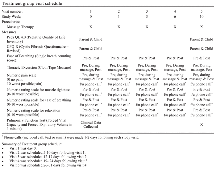
Participants in the treatment group received a series of three to five MT treatments over 10 to 12 weeks. Each treatment lasted 60–90 min. If diagnosed with a pulmonary exacerbation requiring hospitalization, treatment participants were permitted to continue in the study with pulmonary physician approval. Participants with a history of hepatomegaly or splenomegaly were eligible for the study with the provision that abdominal massage was not performed. If the participant had recently received MT, the first study visit was delayed for 30 days.
For all treatment participants, time between visits was incrementally increased, following a common outpatient model for massage therapy. Anecdotally, as a client’s symptoms improve following repeated sessions, time between visits is often increased. For this study, time between visits increased incrementally from 1 week, to 2 weeks, to 3 weeks, to 4 weeks. This study compared findings over time, looking for patterns in timing of musculoskeletal changes following MT and how long the changes lasted.
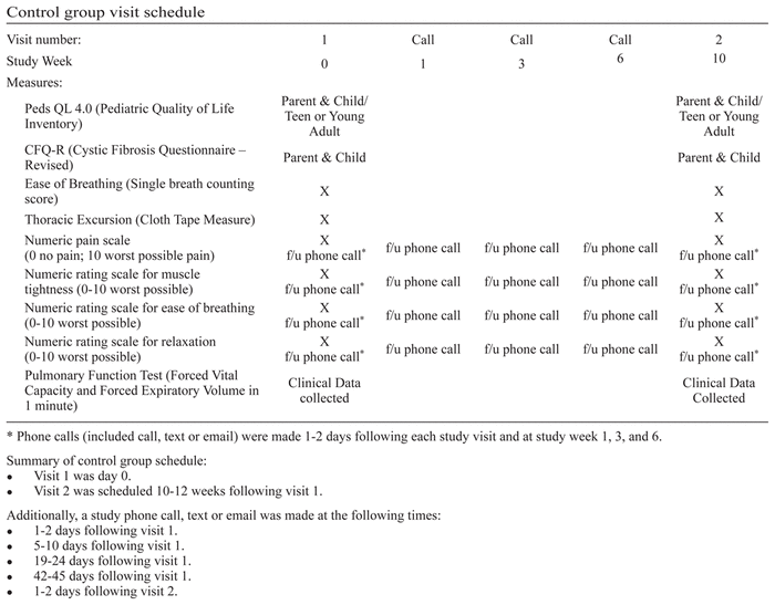
Participants in the control group were seen twice in the outpatient clinic for data collection at study Weeks zero (time one) and 10 (final study visit). Follow-up phone calls correlated with treatment group visits at Weeks One, Three, and Six. The control group did not receive MT during active study participation.
Participation in the study was voluntary; participants could withdraw at any time. The study included financial compensation for study participants (maximum $60 control group; $90 treatment group). At the conclusion of the study, small bolsters used during MT were to be given to the treatment group participants. A courtesy massage was to be offered to the control group participants at completion of data collection.
Data were collected by trained study assistants during and between visits. Massage therapists were trained in, and assisted with, data collection outside of the treatment visit.
Descriptive analyses were performed on sample demographics and outcome variables in the treatment and control groups. Two-sample t test and Fisher’s exact test were applied for demographic comparison. Continuous variables were summarized using means, standard deviations, and ranges, while categorical variables were described using counts and percentages. Final outcome measures between the treatment and control groups were compared by ANCOVA controlling for age, gender, and baseline prescores. Effect size Omega-squared (ω2) and Cohen’s d were calculated to estimate magnitude of the effect. According to Cohen,(13) 0.14, 0.06 and 0.01 are the cut points for ω2 and 0.8, 0.5 and 0.2 for d in terms of large, moderate, and small effects. Data were analyzed employing SAS statistical software, version 9.3 (SAS Institute, Cary, NC). A two-sided significance level was set at 0.05.
The PedsQL, originally developed to measure health related QOL in children ages 2 to 18 years old,(14) was modified to the PedsQL 4.0, a 23-item scale of general health-related quality of life (HRQOL) in children and adolescents and young adults.(15) The PedsQL 4.0 has demonstrated internal consistency reliability for the total scale score (child self-report α = 0.88, parent proxy report α = 0.90), psychosocial scale score (child self-report α = 0.83, parent proxy report α = 0.86), and physical health summary scale score (child self-report α = 0.80, parent proxy report α = 0.88). Construct validity was demonstrated using known groups method.(16) This study used the self-report scales and parent proxy reports for children and teens, and self-reports for young adults.
The CFQ-R is a disease-specific measure of QOL among children and adults with CF.(17) The child version is a 35-item tool with eight domains: physical, emotional state, social, body image, eating, treatment burden, respiratory, and digestion. Each scale yields a standardized score ranging from 0–100, with higher scores indicating better HRQOL.(18) The Adolescent/adult version demonstrates internal consistency (α = 0.67–0.94) and test/retest reliability (rs = 0.45–0.90) among patients with CF ages 14 to 53 years old.(19) The child version demonstrates internal consistency and reliability (α = 0.7–0.92) among children with CF ages 6 to 13 years old.(20) Construct validity has been tested by means of correlation with FEV1 % predicted (r = 0.01–0.18). The parent version demonstrates internal consistency (α = 0.59–0.91) and parent/child agreement = 0.27–0.57.(20) This study focused on the child and teen reports.
In this study, the author created three numeric rating (0–10) scales capturing subjective descriptions of QOL in individuals with CF:
Muscle tightness. Participants were asked: “On a scale from 0 to 10, how would you describe how your muscles feel right now, with 0 being the least tight (you are able to move very easily), and 10 being the most tight, the greatest amount of tightness and greatest difficulty moving you have ever experienced?”
Ease of breathing. Participants were asked: “On a scale from 0 to 10, how would you describe your breathing right now, with 0 being the least difficulty breathing (you are able to breathe very easily), and 10 being the most difficulty breathing you have ever experienced?”
Relaxation. Participants were asked: “On a scale from 0 to 10, how would you describe your overall relaxation right now, with 0 being the least tense and stressed (you feel the most relaxed and calm you have ever felt), and 10 being the most tense and stressed you have ever felt?”
In this study, pain was measured using a previously established scale, the Numeric Rating Scale (NRS-11), which is a self-report measure of pain intensity for children 8 years and older, with endpoints corresponding to no pain (0) and the worst pain possible (10). The pain scores are categorized as mild (1–3), moderate (4–6), and severe (7–10). Previously, validity was demonstrated with three samples of children: correlation with the Faces Pain Scale – Revised (r = 0.87); a visual analogue scale (VAS) (r = 0.89); and between-group comparisons of the distribution of pain intensity scores on the NRS-11 and VAS which showed very similar distributions (paired t = 1.88, df = 117, p = .06), except for scores at the very lowest end of the scales (p = .03 at 0).(21) In this study, the NRS scale was administered at predetermined times to evaluate potential effects of MT on musculoskeletal pain.
Measurements for this study included the Forced Vital Capacity (FVC) and Forced Expiratory Volume in 1 sec (FEV1), performed using the American Thoracic Society standards.(22) Previous studies used pulmonary function measurements in individuals with CF who received MT.(9,10,12) Forced Vital Capacity is the total amount of air forcefully exhaled after a full inspiration. During spirometry, the subject must inhale as much air as possible and then exhale as much air as possible. The “F” in FVC is used because this is a forced maneuver. FEV1 is the total amount of air forcefully exhaled in the first second following a full inspiration.
Assessment of the ease of breathing for this study was measured using the Single Breath Counting Score.(23) This score was determined by having the subject stand with arms relaxed at their sides, take a large breath and then count out loud, reading the numbers from a digital stop watch aloud, one number per second. The highest number the subject was able to verbalize before taking the next breath was the score. The Single Breath Counting Score was previously found to demonstrate test/retest reliability at rest (ICC = 0.93), and construct validity through correlations with FEV1 after exercise (r = 0.68, p < .001), not at rest.
The upper and lower thoracic excursion of each subject was measured by the cloth tape measure technique of the subject’s chest.(24) Both upper and lower thoracic excursion (thoracic circumference) were determined by the following formula: forced inspiration minus forced expiration = thoracic excursion. Test/retest reliability was previously determined with upper thoracic excursion ICC = 0.91 and 0.86 (95% CI, 0.69–0.99) for two sessions of measurement, respectively. Lower thoracic excursion ICC of 0.84 and 0.81 (95% CI 0.69–0.99) was obtained at two measurement sessions respectively.
A specific MT protocol based on deep tissue myofascial TrP therapy(25) was developed by the lead author in collaboration with MT experts. The massage protocol blended Swedish strokes and myofascial TrP therapy, creating a full body sequence of massage strokes. Massage techniques were provided by a licensed massage therapist (LMT) and focused on releasing TrPs by applying pressure to affected tissue for 60–90 sec, followed by stretching techniques, specifically PIMR (post-isometric muscle release), to retrain the affected muscle to stay lengthened to its natural resting state. The massage focused primarily on muscles involved in supporting respiration and posture, areas commonly compromised in individuals with CF. Each treatment group participant was assigned to a specific LMT. When the LMT was unavailable, another study trained LMT provided the massage.
The massage therapy protocol included application of massage strokes with the participant placed in three different positions on a massage table: supine, side-lying and sitting. The sequence of massage techniques were administered over 60–90 min and included:
In the supine position, gentle bilateral leg stretch and TrP release of quadratus plantae; sacro-sternal fascial releases; L5-S1 release; upper trunk and bilateral shoulder warm-up; chest warm-up; digital kneading bilateral temporalis muscles; posterior neck and suboccipital massage; trapezius warm-up and release; serratus anterior release; pectoralis major TrP release; rectus abdominis and psoas TrP release; tensor fascia lata stretch and PIMR; and ended with assistive inspiration.
In the side-lying position (left side down, then repeated all techniques with right side down), spine warm-up with fascial stretches; erector spinae rocking; intercostal stretch and TrP release; levator scapula TrP release and PIMR; rhomboid TrP release and PIMR; and ended with pectoralis minor TrP release and PIMR.
In the sitting position on side of table, thoracic PIMR for external intercostals; rotational trunk PIMR for intervertebral muscles.
The entire protocol ended with resting hands on shoulders followed by relaxation stroking down back.
Three LMTs assisted the Principal Investigator (PI) in study related activities. Two of the LMTs had pediatric hospital experience in caring for hospitalized youth and young adults with CF. The third LMT had extensive experience in myofascial TrP therapy and assisted in development of the study MT protocol. The LMTs were trained in the MT protocol over three practice sessions by the PI and the LMT who assisted in protocol development. Additionally, the PI had extensive experience in providing massage therapy to hospitalized individuals with CF at the study hospital. All LMTs, including the PI, demonstrated the ability to consistently provide the massage protocol as outlined for the study with replicable skills.
In an effort to determine the body’s ability to maintain musculoskeletal changes following the massage protocol, time between treatments was gradually increased. The required three to five MT treatments were administered over 10 to 12 weeks. (See Appendix B: Timeline).
This pilot study enrolled a convenience sample of 30 males and females, 8- to 21-year-olds; 24 participants completed the study and six were lost to attrition (four declined participation after initial enrollment; one was unable to be reached; and one was withdrawn due to missed visits (only completed two of the three required visits). All remaining participants completed the required number of visits/phone data collection. All participants in the study were Caucasian, ranging in age from 9.9 to 20.6 years old, with a mean age of 15.7 (SD = 3.5) years. Sixteen (66.6%) of the participants were female. There were no significant differences for age and gender (p > .66) between the treatment and control groups. The number of hospitalizations was compared between the treatment and control groups. Three participants in the treatment group were hospitalized four times collectively and six participants in the control group were hospitalized 16 times collectively, (ranging from 1 to 8 times). The number of hospitalizations in treatment group was lower (Mean (SD) = 0.33 (0.65) than control group (Mean (SD) = 1.35 (2.30), but did not achieve statistical significance (Wilcoxon rank-sum test, Z = 1.2741, p = .2026) with close to medium effect size (r = 0.24).
Quality of life was measured with the PedsQL 4.0 and CFQ-R and with subjective measures of muscle tightness, ease of breathing, and relaxation created for this study. Previous studies described similar quality of life indicators for individuals with CF (e.g., subjective reports of improved breathing;(9,10,12) decreased pain, less restricted breathing, and increased relaxation(21)); and reduced muscle tightness, easier breathing, increased muscle strength and chest expansion, improved sleep, increased relaxation, and an overall feeling of improved well-being.(10)
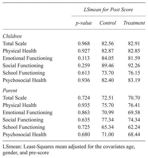
No statistically significant differences in children’s and caregivers’ QOL scores in treatment group between pre- and post-MT intervention or compared to the control group was achieved when using the PedsQL 4.0 and the CFQ-R tools. However, PedsQL 4.0 scores clinically improved in the treatment group in psychosocial ((Mean (SD) = 84.72 (15.04) vs. Mean (SD) = 82.22 (16.43)), physical ((Mean (SD) = 85.94 (18.63) vs. Mean (SD) 83.59 (16.86)), and total health scores ((Mean (SD) = 85.14 (15.66) vs. Mean (SD) = 82.70 (15.73)) with Cohen d of 0.21, 0.08, and 0.25. When compared to the control group, by the end of the study, the child psychosocial and total health subscales in the treatment group improved a little more with very small effect sizes (ω2 = 0.006, and 0.005) (see Table 1). Additionally, the post parent/caregiver physical health scores improved more in the treatment group than the control group (ω2 = 0.010).
Table 1 Children and parent post-intervention Peds QL 4.0 score
The treatment group reported a decrease in muscle tightness ((Mean (SD) = 0.36 (1.81) vs. Mean (SD) = 1.80 (1.48), d = 1.06)); greater ease of breathing ((Mean (SD) = 0.73 (1.01) vs. Mean (SD) = 0.50 (0.97), d = 0.06)); and improved relaxation ((Mean (SD) = 0.91 (1.14) vs. Mean (SD) = 1.40 (1.35), d = 0.66)) at the last follow-up call. Compared to the control group, muscle tightness in the MT group was significantly reduced (p = .048) at post-visit 5, with a large effect size (ω2 = 0.163). Relaxation scores in the MT group were improved (p = .052) compared to the control group at post-visit 5, with a large (ω2 = 0.156) effect size (see Table 2). However, results were not statistically significant. Of note, the male participants had significantly higher PedsQL scores on emotional, social, and school functioning, and general psychosocial health than the female participants at the conclusion of the study. Interestingly, when comparing a child one year younger in age (e.g., 10-year-old compared to 11-year-old), the younger participant had significantly higher social functioning scores at the end of the study (p = .0466).
Table 2 Clinical outcome means and standard deviations, not adjusted for covariates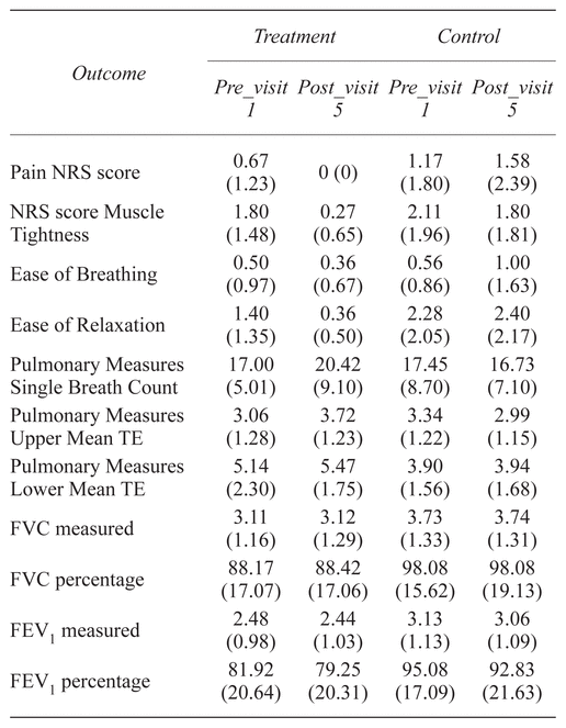
When comparing the participants’ pain scores of mild, moderate, and severe, the treatment group reported lower Pain NRS score ((Mean (SD) = 0 (0) vs. Mean (SD) = 0.67 (1.23), d = 0.94)) at the last follow-up call. NRS pain scores had improved compared to the control group at post-visit 5, with marginal significance (p = .076) and a moderate effect size (ω2 = 0.095) (see Tables 2 and 3).
Table 3 Postintervention Clinical Measurements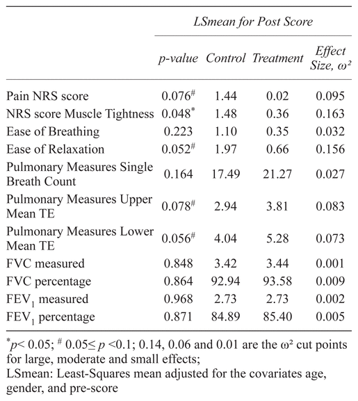
Pulmonary function measures included upper and lower thoracic excursion and spirometry scores (FEV1 and FVC). The upper and lower Mean TE scores in the MT group were improved at the post-visit 5 ((Mean (SD) = 3.72 (1.23) vs. Mean (SD) = 3.06 (1.28), d = 0.53)) and ((Mean (SD) = 5.47 (1.75) vs. Mean (SD) = 5.14 (2.30), d = 0.26)). Compared to the control group, the upper and lower Mean TE scores were marginally statistically significantly higher (p = .078, and p = .056) in the last visit, with moderate effect sizes (ω2 = 0.083 and ω2 = 0.073) (see Table 2). The PFT in the treatment group showed a slightly higher trend than in the control group. The participant who was 1 year older in age (e.g., 10-year-old compared to 9-year-old) (p = .0418) had significantly higher FVC percent predicted measure by 1.13 at the end of the study (see Table 3). The treatment group had higher single breath counting scores (d = 0.47) than the control group at the last study visit. No statistically significant changes in PFTs were found for either group over time.
Cystic Fibrosis is a common, life-shortening genetic disease which may negatively affect QOL. With disease progression, individuals may experience an increase in pulmonary exacerbations including increased work of breathing and related cough, and musculoskeletal changes with pain and tissue dysfunction. Children with mild CF have reported frequent mild pain in chest, head/neck, and abdomen.(6) Previously, youth with CF were found to experience significant decreases in QOL as pain increased or persisted over time.(7)
Additional findings supported the use of massage therapy in individuals with CF to improve QOL—for example, MT significantly decreased pain in hospitalized youth,(8) decreased anxiety and improved mood and peak airflow in children,(12) and decreased muscle tightness and musculoskeletal pain in postural muscles in adults with CF.(9,10) Other subjective descriptions of improved QOL included improved breathing and pulmonary function,(8,9,10) decreased pain, less restricted breathing, and increased relaxation,(26) and reduced muscle tightness, easier breathing, increased muscle strength and chest expansion, improved sleep, increased relaxation, and an overall feeling of improved well-being.(10)
As previously discussed in the Results section of this pilot study, statistically significant results were not achieved for some outcomes with large and moderate effect sizes between the treatment and control groups. These findings may be due to the low statistical power attributed to the small sample size.
This study aimed to explore the trend and effect sizes in the measured outcomes to determine the feasibility and potential value of future large scale multi-site studies. In this sense, effect size and trend were more useful than statistical significance due to the small sample size (N = 24) which could not provide enough power for the statistical tests to detect statistical significance on some large and moderate effect sizes. Number of hospitalizations in treatment group was lower than control group with a close to medium size effect, but did not achieve statistical significance. There were inconsistent findings from the PedsQL and CFQ-R tools. The time-specific subjective measures of QOL, including pain, muscle tightness, ease of breathing, and overall relaxation, found positive effects of MT and may be more sensitive to treatment specific changes. There were no significant changes in PFTs. However, improved ease of breathing and thoracic excursion scores were found in participants who received MT. A study limitation was the applicability of using the single breath counting score for subjects at rest. Test/retest reliability had been previously found at rest, and construct validity found after exercise. Additionally, the data did not include reports of significant life events which could impact QOL.
The feasibility of conducting a study on MT in CF over 10 to 12 weeks was demonstrated in this pilot study. Trends in the data suggested that the use of a myofascial TrP massage in CF care may be beneficial. Overall, the results of this study contributed to the body of knowledge related to the effects of MT on individuals with CF. However, the protocol was based on MT outpatient practices and is limited in its application to outpatient CF care (no related studies found). Future recommendations include a shortened version (15 to 30 min) of the study protocol, using subjective measures of QOL and modifying the deep tissue myofascial TrP protocol to focus on areas where TrPs most frequently occur in this population. This study primarily found TrPs in the intercostal muscles, thoracic erectors, and sternal and lower ribcage attachments. A shortened MT protocol targeting musculoskeletal dysfunction and pain may be more feasible for routine outpatient CF clinic visits (e.g., every three months) than a longer, more frequent MT intervention. Preventative measures in CF care, including MT, may subsequently be found to improve musculoskeletal function and form, decrease related pain (e.g., triggered by cough and increased respiratory effort), and may ultimately contribute to a decrease in frequency of hospitalizations and an increase in quality of life in individuals with CF.
This study was supported by a grant from the Carolyn Stoll Nursing Research Fund, Cincinnati Children’s Hospital Medical Center. This funding source had no specific involvement in the conduct of this research or related presentations, publications or other reporting activities. Earle Timberlake, Registered Massage Therapist and clinical expert in myofascial trigger point massage consulted on this study related to massage protocol development. James Taddeo, BSN, RN, LMT and Margaret Bottenhorn, MA, LMT assisted with data collection and massage therapy when the primary therapist was unavailable. Barbara Giambra, PhD, RN, CPNP, Assistant Professor in the Center for Professional Excellence at CCHMC, assisted in the review of literature. Melissa Liddle, MA, CCLS, CTRS assisted in manuscript development.
Kate Zink dedicates this study to the courageous young individuals with Cystic Fibrosis who have inspired her work and reminded her of the value of living our lives to the fullest. With heartfelt appreciation, she wishes each of you joy and a rich and meaningful life.
The authors declare there are no known conflicts of interest associated with this study.
1 Cystic Fibrosis Foundation Patient Registry. 2016 Annual Data Report. Bethesda, MD: Cystic Fibrosis Foundation; 2007. [Last accessed on 2018 January 16]. Available from: https://www.cff.org/Research/Researcher-Resources/Patient-Registry/2016-Patient-Registry-Annual-Data-Report
2 Hamosh A, FitzSimmons S, Macek M, Knowles M, Rosenstein B, Cutting G. Comparison of the clinical manifestations of cystic fibrosis in black and white patients. J Pediatr. 1998;132(2):255–259.

3 Elborn, S. Cystic fibrosis. The Lancet. 2016;388(10059): 2519–2531.
4 Travell, J, Simons D. Myofascial Pain and Dysfunction: the Trigger Point Manual Vol II. Baltimore, MD: Lippincott Williams and Wilkins; 1992.
5 Simons D, Travell J, Simons L. Myofascial Pain and Dysfunction: the Trigger Point Manual Vol I, 2nd ed. Baltimore, MD: Lippincott Williams and Wilkins; 1999.
6 Koh J, Harrison D, Palermo T, Turner H, McGraw T. Assessment of acute and chronic pain symptoms in children with cystic fibrosis. Pediatr Pulmonol. 2005;40(4):330–335.
7 Palermo T, Harrison D, Koh J. Effect of disease-related pain on the health-related quality of life of children and adolescents with cystic fibrosis. Clin J Pain. 2006;22(6):532–537.
8 Zimmer M, Bogenschutz L, Zink K. Effect of Massage Therapy on Pain in Hospitalized Pediatric Cystic Fibrosis Patients [abstract]. North American Research Conference on Complementary and Integrative Medicine, 2008 [cited 2009]. [Last accessed on 2018 January 31]. Available from: https://clinicaltrials.gov/ct2/show/NCT01729585
9 Lee A, Holdsworth M, Holland A, Button B. The immediate effect of musculoskeletal physiotherapy techniques and massage on pain and ease of breathing in adults with cystic fibrosis. J Cyst Fibros. 2009;8(1):79–81.
10 McQueen K, Button B, Heathcote C. Massage and musculoskeletal physiotherapy service for adults: Effects on chest mobility, posture, muscle tension, pain, ease of breathing and well-being. Pediatr Pulmonol. 2003;Supplement 25(266):240.
11 Field T, Henteleff T, Hernandez-Reif M, Martinez E, Mavunda K, Kuhn C, et al. Children with asthma have improved pulmonary functions after massage therapy. J Pediatr. 1998;132(5):854–858.
12 Hernandez-Reif M, Field T, Krasnegor J, Martinez E, Schwartzman M, Mavunda K. Children with cystic fibrosis benefit from massage therapy. J Pediatr Psychol. 1999;24(2):175–181.
13 Cohen J. Statistical Power Analysis for the Behavioral Sciences, 2nd edition. Hillsdale, NJ: Lawrence Earlbaum Associates; 1988.
14 Varni J, Seid M, Rode C. The PedsQL: measurement model for the Pediatric Quality of Life Inventory. Med Care. 1999;37:126–139.
15 Varni J, Seid M. Kurtin P. PedsQL™ 4.0: Reliability and validity of the Pediatric Quality of Life Inventory version 4.0 Generic Core Scales in healthy and patient populations. Med Care. 2001;39(8):800–812.
16 Varni J, Burwinkle T, Seid M, Skarr D. The PedsQL™ 4.0 as a pediatric population health measure: feasibility, reliability, and validity. Ambul Pediatr. 2003;3(6):329–341.
17 Henry B, Aussage P, Grosskopf C, Goehrs JM, Launois R, French CFQOL Study Group. Cystic Fibrosis Questionnaire—Revised. New York: Am Thoracic Society; 2002. Available from: http://qol.thoracic.org/sections/instruments/ae/pages/cfq-cfq-r.html
18 Modi A, Quittner A. Validation of a disease-specific measure of health-related quality of life for children with cystic fibrosis. J Pediatr Psychol. 2003;28(8):535–545.
19 Quittner A, Buu A, Messer M., Modi A, Watrous M. Development and validation of the Cystic Fibrosis Questionnaire in the United States: a health-related quality-of-life measure for cystic fibrosis. Chest. 2005;128(4):2347–2354.
20 Quittner A, Modi A, Cruz I. Systematic review of health-related quality of life measures for children with respiratory conditions. Pediatr Resp Rev. 2008;9(3):220–232.
21 Von Baeyer C, Spagrud L, McCormick J, Choo E, Neville K, Connelly M. Three new datasets supporting use of the Numerical Rating Scale (NRS-11) for children’s self-reports of pain intensity. Pain. 2009;143(3):223–227.
22 Miller M, Hankinson J, Brusasco V, Burgos F, Casaburi R, Coates A, et al. Standardisation of spirometry. Eur Respir J. 2005;26(2):319–338.
23 Orenstein D, Holt L, Rebovich P, Campbell T, Nixon P. Measuring ease of breathing in young patients with cystic fibrosis. Pediatr Pulmonol. 2002;34(6):473–477.
24 Bockenhauer S, Chen H, Julliard K, Weedon J. Measuring thoracic excursion: reliability of the cloth tape measure technique. J Am Osteopath Assoc. 2007;107(5):191–196.
25 Timberlake E. Somatic Release in Clinical Massage, rev. edition. Logan, OH: Timberlake Massage Therapies; 1999.
26 Kemper K, McLellan M, Highfield E. Massage therapy and acupuncture for children with chronic pulmonary disease. Clin Pulm Med. 2004;11(4):242–25
The techniques and sequence detailed below were performed at each study visit. Techniques included variability in strokes, amount of pressure and frequency, e.g., repeated 3–5 times, per therapist’s professional judgment, to achieve muscle release. Flaxseed pillows and standard pillows or half round bolsters were used for proper positioning.
Supine position. Bolsters at occiput and under knees.
Gentle leg stretch followed by TrP (trigger point) release of quadratus plantae at soles of feet and Post Isometric Muscle Release (PIMR) each leg.
Sacro-sternal fascial releases. Hold sacrum in palm of hand and allow tissue to relax. Apply gentle pressure with other hand on abdomen, lower ribcage, and upper ribcage/sternum and traction toward umbilicus.
L5-S1 release. Stand on right side, place open palm right hand under sacrum and open fisted left hand horizontal at L5, cupping spinous processes. Gently distract hands away from one another.
Upper trunk/shoulder warm up, right and left sides. (Bolster under shoulder being worked) Passive arm stretches repeated 5–10 times in three arm positions:
Upper arm at 90 degrees, elbow flexed, palm down. Hand raised toward head, then down to side.
Upper arm at 90 degrees, elbow flexed, palm down. Elbow raised toward head and back down to 90 degree flexion.
Upper arm at 90 degrees, palm facing up. Arm raised over head, fingers extended. Return arm to 90 degrees flexion.
Remove bolster, reposition under left arm/shoulder and repeat sequence on left side. Remove bolster.
Chest warm up. Digital kneading bilaterally, sterno-costal points of attachment, from clavicle to xiphoid process (x3). Apply gentle pressure to points of attachment bilaterally and perform horizontal tissue glide (x3). Perform costal raking upper chest bilaterally, medial to lateral (5–10 times).
Temporalis. (Remove bolster from under head). Gentle digital kneading bilateral temporalis muscles 15–30 seconds.
Posterior neck and sub occipitals. Cradle head with fingertips on posterior cervical muscles.
With fingertips bilateral to cervical muscles, apply gentle medial pressure alternating left to right from upper to lower cervicals (x3).
Trapezius warm up and release. Cradle occiput with left hand while applying pressure with right thumb to mid sagittal suture on top of head × 1–2 minutes. Sift trapezius for areas of restriction and release 1–2 TrP’s as available. Sift scalenes and release Trp’s as indicated. Lift and cradle head on open palms, head in forward flexion, × 1–2 minutes. Follow with occipital base release (head on table). Right hand on left shoulder and left hand cradling head on left, gently resist lateral head flexion left while gently resisting left shoulder shrug up and perform PIMR × 1. Left hand on right shoulder and right hand cradling head on right, gently resist lateral head flexion right while gently resisting right shoulder shrug up and perform PIMR × 1. With therapist’s arms crossed and under subject’s head, subject’s eyes open, move head up to barrier. Instruct subject to push back into therapist’s arms, breathe in, and then relax head and allow therapist to move head farther into flexion. Invite subject to drop shoulders down to enhance the stretch.
Serratus anterior. (Bolster under head). Right side. Locate TrP at T6 on lateral rib. Instruct subject to breathe in while therapist holds TrP with finger tips, raising subject’s hand above head and lowering flexed arm to side. Repeat sequence as indicated. Finish with PIMR × 1.
Pectoralis major. Bolster under right elbow, arm flexed 90 degrees with palm on table. Sift lateral body of pectoralis major, searching for 2–3 trigger points and release if found. Perform pectoralis major stretches with subject close to side/edge of table. Stretch at 90 degrees abduction (palm up), opposite hand fixed on the sternal head (between breast tissue) followed by PIMR × 3.
Repeat above sequence (serratus anterior and pectoralis major) on left side.
Rectus abdominis and psoas (omit this section for persons with hepatomegaly or splenomegaly). Starting on left side, flex knees, feet flat on table. With finger tips, follow inferior costal border medial to lateral, for 2 inches, searching for TrP’s; hold 60 seconds. Allow left flexed knee to drop laterally, externally rotating hip. Search anterior ileum for TrP’s and release, including ASIS joint. Right leg on table, flex left leg 90 degrees off table. Gently press fingertips into abdomen 1–2” lateral to umbilicus while subject lifts flexed leg and therapist locates psoas muscle. Apply light to moderate pressure, allowing psoas to soften (30–60 seconds). Release pressure. Perform PIMR flexed leg × 1.
Repeat rectus abdominis and psoas releases on right side.
Tensor fascia lata (TFL). Therapist on right side. Left leg extended, right leg flexed, foot crossed left knee and foot flat on table. With left hand, gently push right knee to left while pulling left ankle with right hand across midline. Instruct subject to pull left leg lateral (to the left) and push right knee into therapist’s hand (to the right) to perform PIMR × 3; end last PIMR with medial pull/jerk. Repeat on left side, changing direction of PIMR stretches accordingly. Remove bolster from head.
Assistive inspiration. Small bolster under lower back. Therapist positioned at head of table with subject raising arms up with palms toward ceiling. Therapist grasps hands, palm to palm, interlocking thumbs and bringing arms superior, overhead with subject inhaling, to expand chest. Subject instructed to exhale while therapist brings arms down toward abdomen and therapist applies gentle downward pressure on inferior ribcage; perform X 3.
Side lying position, left side down. Small bolster under waist to arch spine laterally; large bolster anterior to chest for arm support and between legs/knees for leg support; pillow bolster under head.
Spine warm up. Standing posterior to subject, perform passive touch and gentle back stretch; one hand on sacrum, other hand on upper back, stretching fasciae, hands away from one another.
Erector spinae. Apply thumb presses with rocking motion along spine (deep paraspinals), from upper neck to sacrum, on the up side of vertebrae, X 3.
Intercostals. Apply gentle stretch, one palm down and other palm on top of hand. Gently press fascia inward and anterior, over ribs. Upper arm above head, subject’s hand holding head of massage table. Using P-ball or fingertips, apply gentle intercostal presses from T4 (under axilla) to T12, posterior to anterior. Treat costal angle going anterior, and then treat anterior intercostals under breast.
Levator scapula. Perform digital kneading of levator scapulae with upper arm over head. With fingertips, locate TrP’s at insertion of levator scapulae at superior border and medial angle scapula. Treat TrP with massage tool at angle of neck where muscle surfaces, at muscle twist at anterior border of trapezius and superior medial angle of scapula.
Rhomboids. Position upper arm anterior to subject, closed fist on massage table and elbow flexed and pointed toward ceiling. Search rhomboids inferior to superior (cross fiber) and treat myofascial trigger points along vertebral border of scapula. Move upper arm in extension above head and back to 90 degree flexion. Finish with PIMR rhomboids × 1 by extending arm over head (pushing anterior at shoulder).
Pectoralis Minor. With upper arm relaxed across body and lying on large bolster, palm down, perform myofascial TrP therapy. Work with fingertips/thumb deep to lateral body of pectoralis major from attachments on 5th, 4th, and 3rd ribs toward coracoid process. Perform PIMR × 1. Assist subject in positioning to the right side; repeat above steps 2–6.
Sitting position. Therapist assists subject to sitting position on side of the table.
Thoracic PIMR for external intercostals. Therapist in front of subject, subject takes breath in while therapist gently assists subject in performing a side-bending movement right, at which time subject releases the breath and therapist extends the stretch with gentle pressure on right shoulder. Perform PIMR X 1. Therapist returns subject to center position and repeats sequence left.
Rotational trunk PIMR for intervertebral muscles. Therapist behind subject, instructs subject to cross arms at chest, hands on opposite shoulders.
First position – therapist rotates subject to left (grasping right elbow), while subject turns head right. Therapist instructs subject to take breath in × 5 seconds and release the breath out while therapist continues to gently rotate trunk left with head turning left. Therapist supports back with opposite hand.
Second position – therapist rotates subject to right (grasping left elbow), while subject turns head left. Therapist instructs subject to take breath in × 5 seconds and release the breath out while therapist continues to gently rotate trunk right with head turning right. Therapist supports back with opposite hand.
Resting hands on shoulders. Therapist gently rests hands on subject’s shoulders × 15–30 seconds.
Relaxation stroking. To finish treatment, therapist uses full open hands palms down to stroke subject from the top of the head, down the full back. Perform X 3.
Published under the CreativeCommons Attribution-NonCommercial-NoDerivs 3.0 License. ( Return to Text )
INTERNATIONAL JOURNAL OF THERAPEUTIC MASSAGE AND BODYWORK, VOLUME 12, NUMBER 1, MARCH 2019