Erin E. Cubick , PT, DPT, LAT, ATC, CSCS , Vanessa Y. Quezada , PT, DPT , Ariel D. Schumer , PT, DPT , Carol M. Davis , PT, DPT, EdD, MS, FAPTA
Department of Physical Therapy, University of Miami, Coral Gables, FL, USA.Background:
Myofascial release (MFR) is a manual therapeutic technique used to release fascial restrictions, which may cause neuromusculoskeletal and systemic pathology.
Purpose:
This case report describes the use of sustained release MFR techniques in a patient with a primary diagnosis of rheumatoid arthritis (RA) and a secondary diagnosis of collagenous colitis. Changes in pain, cervical range of motion, fatigue, and gastrointestinal tract function, as well as the impact of RA on daily activities, were assessed.
Methods:
A 54-year-old white woman presented with signs and symptoms attributed to RA and collagenous colitis. Pre and post measurements were taken with each treatment and during the interim between the initial and final treatment series. The patient recorded changes in pain, fatigue, gastrointestinal tract function, and quality of life. Cervical range of motion was assessed. Six sustained release MFR treatment sessions were provided over a 2-week period. Following an 8-week interim, two more treatments were performed.
Results:
The patient showed improvements in pain, fatigue, gastrointestinal tract function, cervical range of motion, and quality of life following the initial treatment series of six sessions. The patient maintained positive gains for 5 weeks following the final treatment, after which her symptoms returned to near baseline measurements. Following two more treatments, positive gains were achieved once again.
Conclusions:
In a patient with RA and collagenous colitis, the application of sustained release MFR techniques in addition to standard medical treatment may provide short-term and long-term improvements in comorbid symptoms and overall quality of life.
KEYWORDS: Myofascial release , rheumatoid arthritis , inflammatory bowel disease , manual therapy
Rheumatoid arthritis (RA) is a chronic, systemic, polyarticular inflammatory disease, causing destruction and inflammation to the capsule and synovial lining of joints throughout the body.(1,2) The local inflammatory injury is induced by pathological immune complexes traveling through the circulatory system.(1) T-lymphocytes, leukocytes, monocytes, and cytokines infiltrate the synovial fluid causing joint inflammation.(1) Tumor necrosis factor, a primary cytokine in RA, stimulates protein-degrading enzymes to be released.(1) This results in destruction of articular cartilage, bone resorption, and inhibition of bone formation.(1) Another pathological immune complex, rheumatoid factor, is an antibody that may trigger inflammatory reactions if it interacts with immunoglobulin antibodies and the mediators of inflammation.(1,3) This reaction, along with specialized effector cells, causes pannus tissue, a destructive vascular granulation tissue, to be formed.(1) As a result, degraded cartilage and bone, effusion of the joint capsule, and thickening of the capsule’s synovial lining occur.(1,3) These changes prevent the synovial joints from being lubricated and providing nutrition to the articular cartilage.(1) Pain, swelling, and gradual destruction of the joint can result in loss of function, deformity, and, ultimately, disability.(1,3)
RA usually develops in the third or fourth decade of life, affecting three times more women than men.(1,2) The autoimmune inflammatory disease primarily affects the synovial joints, especially the knees, hands, and feet.(2) The progressive development of RA can lead to prolonged morning stiffness, limited range of motion (ROM), symmetrical joint deformity, ligamentous laxity, altered biomechanics and posture, rheumatoid nodules, pain, fatigue, malaise, fever, weight loss, neurological compromise, and decreased quality of life.(1,2,4,5) As it is an autoimmune disorder, the cardiovascular, pulmonary, and gastrointestinal (GI) systems may also be affected.(1) This chronic, progressive disease goes through periods of remission and exacerbation.(1) Understanding the pathophysiology of RA has led to various treatment strategies including physical therapy, anti-inflammatory medications, disease-modifying antirheumatic drugs, immunosuppressants, and surgery.(4) Sustained release myofascial release (MFR) is also reported to be a valuable healing catalyst that may enable improvements in these realms for individuals with RA.(6)
Individuals with RA may have an excess of human tumor necrosis factor-alpha (TNF-α) causing the body’s immune system to attack healthy tissue. (7) Treatment options for individuals with excess TNF-α include administering anti-TNF antibodies to reduce inflammation and joint damage.(2) Anti-TNF therapy assists in the treatment of RA by restoring the patient’s immune cells and hemoglobin, and reducing the patient’s rheumatoid factor levels, cytokine production, vascular endothelial growth factor, acute-phase protein production, elevated platelets, and elevated fibrinogen.(2)
Infliximab infusion (Remicade) is one type of anti-TNF therapy for patients with moderate to severely active RA.(7) Infliximab binds to TNF-α, interfering with endogenous TNF-α activity.(7) Specifically, this interference prevents inflammatory cytokine and tissue-degrading enzyme induction, leukocyte migration, and neutrophil and eosinophil activation.(7) Thus, infliximab reduces the infiltration of inflammatory cells and mediates cellular adhesions.(7) These actions may prevent RA-associated joint disease and allow diseased joints to heal.(7) Maintenance dosing occurs at 4- to 8-week intervals, depending on the individual’s response to the medication.(7)
Another antirheumatic treatment is methotrexate, an immunosuppressant that inhibits the production of T cells.(2) Methotrexate is considered one of the most effective medications for RA, as it decreases pain and morning stiffness.(1) An effective treatment for RA has been the combination of infliximab with methotrexate, creating a synergistic effect.(2) This combination has been shown to stop cartilage and bone damage in 50% of patients after 6 months of treatment, with effects lasting for 2 years.(2)
Inflammatory bowel disease (IBD) is a chronic inflammatory disease of the GI tract that is classified as a functional disorder of motility in the small and large intestines. Common IBDs include ulcerative colitis and Crohn’s disease, while less common diagnoses include forms of microscopic colitis such as collagenous colitis and lymphocytic colitis.(8) Ulcerative colitis displays chronic inflammation in the mucosa and submucosa of the colon, whereas chronic inflammation from Crohn’s disease can affect any part of the intestinal tract.(1) Microscopic colitis is a chronic inflammatory disease of the colon that causes chronic watery diarrhea.(9) Collagenous colitis causes thickened subepithelial collagen in the colon, whereas lymphocytic colitis has an increase in intraepithelial lymphocytes in the colorectal mucosa.(8,9)
Though the etiology is unknown, IBD may result from genetic predisposition, environmental factors, immunologic factors, chronic stress, inflammation, and/or infection.(1,10) While both genders are equally affected by most IBD forms, collagenous colitis is predominantly found in females.(1,8,9) Common signs and symptoms include abdominal pain, constipation, diarrhea, rectal bleeding, decreased appetite, nausea, vomiting, and weight loss.(10) It is also suggested that IBD may share pathogenic pathways with other autoimmune diseases;(1,8,11) 40% of individuals with collagenous colitis also have autoimmune diseases such as RA.(12,13) Extraintestinal signs and symptoms signifying coexistent IBD and autoimmune diseases include chronic fatigue, fever, night sweats, skin lesions, uveitis, arthritis, migratory arthralgias, and hip pain.(1,10)
IBD displays periods of exacerbation and remission.(1) The signs and symptoms of IBD during these exacerbation periods can negatively affect a person’s quality of life. Current treatment options for IBD include relieving abdominal pain, stabilizing motility, altering diet and nutrition, and altering lifestyle habits.(1) Pharmacological intervention has included antidiarrheals, antispasmodics, immune modifiers, antibiotics, corticosteroids, and aminosalicylates.(1) Similar to the treatment of RA, methotrexate and cytokine-based medications, such as infliximab, are being used to control inflammation in patients with IBD.(1,2,7) Little research has been conducted on the positive effects of combined pharmacological and complementary therapies and on the outcomes of using complementary therapies on the somatic symptoms of IBD.
The fascial system is a protective three-dimensional web matrix of connective tissue that envelops every muscle, organ, gland, and cell in the body, also surrounding the circulatory system, nervous system, musculoskeletal system, and digestive tract to affect the overall shape of the body.(14) There are 12 different fascias or connective tissues in the body, each with varying concentrations of collagen, elastin, and ground substance.(15) Collagen provides support, shape, and stability; elastin allows for flexibility; ground substance cushions every cell.(16) The presence of smooth muscle cells within fascia, along with widespread presence of myelinated and unmyelinated sensory and motor nerve fibers and capillaries, has led to a hypothesis that fascia is an actively adapting organ with functional importance, rather than a passive structural organ alone.(14) Fascia may contribute by assisting in support, protection, cellular respiration, elimination, metabolism, and fluid and lymphatic flow.(17) As a result, physiological and mechanical trauma to fascia at the cellular level may have an effect on posture, cellular health, and the immune system.(6,17–19) There is an important physiological interaction between fascia and the extracellular matrix on the one hand and various cells of the body on the other.(17) “Without the oscillatory activity of the matrix and parenchymal cells, [cellular] metabolism would be stunted or nonexistent.”(17)
It is hypothesized that fascial restrictions occur when fascia reorganizes itself in response to tension and stress.(6,20,21) Fascial restrictions constrict the tissue that is embedded within it, pulling on bones, tendons, and ligaments in an attempt to protect the body from further damage.(6,20–22) As a result, cellular function is disrupted and tremendous strain is placed on soft tissue, as demonstrated by injury, stress, surgical adhesions, scars, inflammatory processes, and anatomical malalignments.(6,18,20–22)
Mechanistic, also referred to as traditional or osteopathic, MFR is used most often synonymously with soft tissue mobilization where the “individual ‘stroke’ or technique on a particular spot of tissue is between a few seconds and 1 ½ minutes.”(21,23,24) Sustained release MFR as taught by Barnes emphasizes the intention to practice sustained pressure and traction over the fascial restriction for a minimum of 3–5 minutes to facilitate the piezoelectric effect to the crystal matrix of fascia.(16,20,21,25) When ground substance in a fascial restriction becomes more solid and less fluid, the piezoelectric effect is stifled and the energetic flow is impeded.(16,25) Electrical impulses are generated in the collagen by compressive and distraction forces within the musculoskeletal system.(20,25) These impulses trigger a cascade of cellular, biomechanical, neural, and extracellular events as the body adapts to external stress.(20,25) In response to internal stress, components of the extracellular fluid change in polarity and charge, affecting fascial motion.(20,25) The stimulation of the gel ground substance of the fascia requires this sustained pressure over time in order to bring about the “melting” of the colloidal part of the tissue and to stimulate a sustained piezoelectric flow of electrons along the tissue, thus maximizing the “energy” flow to the tissue over a longer period of time.(6,25,26) Further, with the extracellular matrix softening and the fascial restriction releasing, pressure on pain-sensitive tissue is relieved and the tissues are rehydrated to allow for conduction of flow of photons and vibration.(25,27) However, Schleip(24) reports that any change in the tissue felt under the hands cannot be attributed either to a softening of the ground substance, termed thixotropy, or to a response to the electron flow from the piezoelectric effect as described by Oschman(16) because of laboratory studies of time and force dependency of connective tissue plasticity.(21,28,29) Schleip suggests that fascial plasticity may be due to the self-regulatory qualities of the client’s nervous system.(24) Health care providers have used MFR in this way in the clinical setting for many years, but evidence beyond case reports, such as randomized and case–control clinical trials, is lacking.
The presentation of combined symptoms of RA and IBD has led to the investigation of further potential therapeutic options. Complementary therapies, such as MFR, are designed to restore homeostasis by relieving restrictions that impede energetic flow, work with the body’s own healing mechanism, and have the possibility of being an effective adjunct to allopathic treatments.(6) This case report describes the changes in ROM, pain, fatigue, and GI tract function following the use of MFR techniques on a patient with a primary diagnosis of RA and a secondary diagnosis of collagenous colitis. The guiding questions were as follows: Does MFR, administered by three practitioners in six 45-minute treatment sessions over a period of 2 weeks, affect cervical ROM and reported pain, fatigue, GI tract function, and quality of life in a patient with RA and collagenous colitis? How long could these changes potentially persist following the last treatment?
A 54-year-old white woman presented with cervical pain, systemic pain, fatigue, and explosive diarrhea due to a primary diagnosis of RA and a secondary diagnosis of collagenous colitis. Over the past 8 years she received infliximab infusions and chiropractic adjustments to address the cervical and systemic pain. None of these treatments resulted in sustained relief longer than a few days. Five years after she started receiving infliximab infusions, collagenous colitis was diagnosed. Her gastroenterologist was astounded that the collagenous colitis presented after receiving infliximab infusions for several years, as that pharmacological treatment is used to control inflammation with IBD. During the course of this study, the patient did not receive chiropractic care, only infliximab infusions.
The patient reported a dull ache in the right elbow, neck, and both temporomandibular joints. She stated that both knees, especially the left, felt achy. As a result of her knee pain, she was unable to wear high heels. She expressed frustration, as she was unable to run or rollerblade and was restricted to a limited amount of walking. She was able to swim the breast-stroke as long as her neck was not in pain. Basic activities of daily living were adversely affected, including exiting the bathtub from a long-sitting position. She noted difficulty with intricate needlework for crocheting and was unable to complete computer-related tasks due to bilateral wrist pain. She reported numbness in her right third and fourth fingers and a flexion contracture of the left third finger. She napped on a daily basis, with an overall feeling of fatigue. Her “normal” GI tract function consisted of nausea, bowel urgency, and explosive diarrhea, but she did not experience this on the day of the initial interview.
The postural assessment revealed slight head rotation to the right, slight elevation of the right shoulder, increased lordosis of the lumbar spine, and anterior rotation of the left ileum. Cervical disk degeneration was noted through x-ray imaging. She had full hip and shoulder ROM, but cervical ROM was limited in all directions.
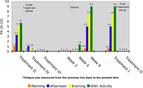
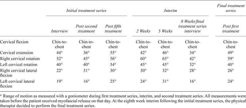
Preceding the first treatment, the patient rated cervical pain as 3.6/10 and systemic pain as 4.8/10. Cervical ROM values were as follows: flexion: chin-to-chest (chin-to-chest was considered full flexion); extension: 44°; right rotation: 52°; left rotation: 40°; right lateral flexion: 22°; and left lateral flexion: 19° (Table 1). She rated her fatigue as 1.1/10 in the morning, 3.3/10 in the afternoon, 5.1/10 in the evening, and 5.7/10 with activity (Figure 1). She scored 164 out of a possible 433 on the Arthritis Impact Measurement Scales 2 (AIMS2; Appendix Table 1).
Table 1
Summary of Cervical Range of Motiona
Appendix Table 1.
Progression of Arthritis Impact Measurement Scales 2 (AIMS2) Scoresa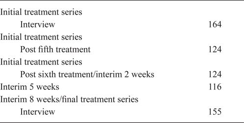
|
|
||
|
Figure 1 Fatigue (P4): comparison of morning, afternoon, evening, and activity fatigue. Overall fatigue was alleviated with myofascial release by the last treatment session of the initial treatment series. The fatigue started to return 5 weeks after the last treatment series and was more severe by 8 weeks. After the final treatment series, the fatigue was eliminated again. |
||
To measure progress, data collected at each treatment and at periodic intervals between treatments included cervical ROM (goniometry), cervical and systemic pain (visual analog scale), and fatigue (P4 Instrument).(30) GI tract function was revealed through patient report. The AIMS2 and the Complementary and Alternative Medicine (CAM) Methods Questionnaire were provided(31) (personal communication, C Ritenbaugh, PhD, MPH, June 28, 2009).
The P4 Instrument was originally created to capture changes in pain throughout the day. However, for the purpose of this study, the original instrument was adapted to capture changes in fatigue. In the adapted version, the word “fatigue” replaced “pain.” The patient provided data on morning, afternoon, evening, and activity-related fatigue. The questions were answered by placing a mark along a visual analogue scale from zero to 10, signifying none to worst possible fatigue.(30)
The AIMS2 was designed to assess the impact of arthritis on daily life. Items assessed included physical function, emotional status, and overall arthritis impact on daily life in the past month. Possible scores ranged from 74 to 433; lower scores indicated wellness and higher scores indicated greater impact of arthritis upon daily life.(31)
The CAM Methods Questionnaire, in a trial phase during the study, was designed to help understand the patient’s experiences throughout in the study. Written quotes were used as well as checked items to quantify changes over time (personal communication, C Ritenbaugh, PhD, MPH, June 28, 2009).
It is hypothesized that MFR is designed to provide more space in the fascia, thereby attempting to relieve pressure on pain-sensitive tissue and improve flow of immune system components, lymphatic fluid, blood, neurotransmitters, neuropeptides, steroids, and digestive enzymes.(16) The MFR technique begins with a postural examination and palpation for fascial restrictions.(26) Then, gentle sustained pressure is used with the hands placed on the restricted tissue, in the direction of the restriction.(26) The pressure is sustained over the skin, without allowing the hands to slide, for a minimum of 90–120 seconds to allow the tissue to begin to release.(26) Once the release begins, pressure is maintained while the therapists’ hands follow the direction of the fascial release.(26) Sustained releases are held for a minimum of 3–5 minutes (Table 2).(26)
Table 2
Summary of a Typical Sustained Release Myofascial Release Treatment Sessiona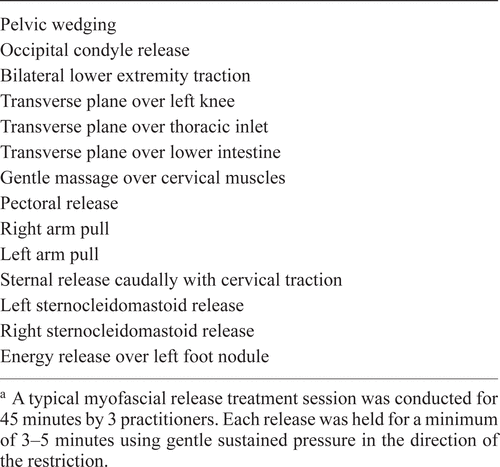
The purpose of this treatment approach was to potentially increase energy levels, decrease pain, and stabilize GI motility to enhance the overall quality of life. Six initial treatments were performed in 2 weeks’ time, followed by an interim of 8 weeks, after which a final treatment series of two sessions was provided. Treatment sessions were provided by a physical therapist experienced in MFR techniques and two trained assistants. Each session was performed for 45 minutes and consisted of several techniques, including manual cervical traction, transverse plane releases, arm and leg pulls, and cross-hand techniques.(26)
The following summary describes changes in signs and symptoms over time. Data collected from each treatment were compared with the most recent values. Post treatment is defined as the timeframe from the conclusion of the last treatment session to the initiation of the next treatment session.
Post first treatment: stabilization in GI tract function (Table 3), systemic pain (Figure 2), and cervical pain (Figure 3). In the 2 days following the first MFR treatment, the patient reported less fatigue than at any other time that she could remember in the past 8 years. Her pain had decreased such that she did not consider taking pain medications in the morning and was able to work at the computer for more than 1 hour. She reported improved sleep at night and was once again able to wear high heels without left knee pain. GI tract function remained stable, as she did not experience any episodes of diarrhea.
Table 3
Summary of Subjective Gastrointestinal Reporta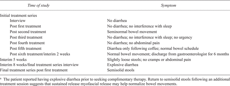
|
|
||
|
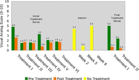
Figure 2 Systemic pain (visual analog scale): comparison of pre and post treatment. Systemic pain was reduced during the initial treatment series and returned during the interim period between the initial and final treatment series. Pain was reduced once again in the final treatment series. |
||
|
|
||
|
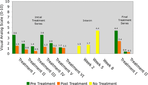
Figure 3 Cervical pain (visual analog scale): comparison of pre and post treatment. The graph depicts a decrease in cervical pain from before each treatment to immediately after each treatment. Cervical pain decreased in the initial treatment series and remained at approximately the same level through the fifth week after the last treatment session of the initial treatment series. Pain was decreased again following one additional myofascial release treatment. |
||
Post second treatment: improved GI tract function (Table 3) and cervical pain (Figure 3); varied cervical ROM (Table 1). Following the second treatment session, she stated she was able to walk for at least 20 minutes pain-free. GI tract function continued to improve based on her subjective report of no bouts of diarrhea or urgency.
Post third treatment: improved GI tract function (Table 3), systemic pain (Figure 2), and cervical pain (Figure 3). On the day of the third MFR session, she received an infliximab infusion. She noted that GI tract function had improved, and she was able to eat all foods without having urgency or diarrhea. She noted that she felt her posture was “straighter” and that her “head [sat] on top of [her] shoulders.”
Post fourth treatment: improved GI tract function (Table 3), systemic pain (Figure 2), cervical pain (Figure 3), and fatigue (Figure 1). She was “astounded by the effects of MFR,” and expressed again that she hadn’t felt this energized in years. At the end of her session, her cervical and systemic pain had decreased to 1.3/10 and 0.8/10, respectively.
Post fifth treatment: improved GI tract function (Table 3), systemic pain (Figure 2), cervical pain (Figure 3), cervical ROM (Table 1), AIMS2 (Appendix Table 1), and quality of life. She noted her fatigue level reached zero. She no longer napped during the afternoons, nor did she use an alarm clock. All GI tract symptoms were eliminated, except for urgency following consumption of coffee. The AIMS2 score decreased from 164 to 124. In the CAM Methods Questionnaire, she recorded a change in energy from 4/10 prior to the study to currently 10/10 and stated that she “felt [herself] coming back to life now with more energy.” She also recorded changes in physical health (pre: 5/10, post: 8.5/10) and happiness (pre: 8.5/10, post: 10/10).
Post sixth treatment; interim, 2 weeks following initial treatment series: improvement maintained in GI tract function (Table 3), systemic pain (Figure 2), cervical pain (Figure 3), and fatigue (Figure 1); maintenance of AIMS2 (Appendix Table 1) and quality of life; varied cervical ROM (Table 1). She returned for an examination 2 weeks following her last treatment to reveal that many of the positive results had been maintained. Her gastroenterologist discharged her for the next 6 months due to regular bowel movements. She recorded changes in energy (pre: 2.5/10, post: 10/10), physical health (pre: 7/10, post: 8/10), and happiness (pre: 10/10, post: 10/10). She felt she was “on a path towards health and wellness” and that her “life [was] getting back to normal.”
Interim, 5 weeks following initial treatment series: improved cervical ROM (Table 1), AIMS2 (Appendix Table 1), and quality of life; regression in GI tract function (Table 3), systemic pain (Figure 2), and cervical pain (Figure 3). The AIMS2 score decreased from 124 to 116. Changes in energy (pre: 1.5/10, post: 10/10), physical health (pre: 5/10, post: 9/10), and happiness (pre: 7.5/10, post: 9.5/10) were recorded through the CAM Methods Questionnaire. She stated that “the fatigue is gone!” and was “using [her] inner resources to heal [herself].” GI tract function maintained relative stability, with reports of slightly loose stools, which may have been a result of a change in daily vitamin intake.
Interim, 8 weeks following initial treatment series: regression in GI tract function (Table 3), systemic pain (Figure 2), cervical pain (Figure 3), cervical ROM (Table 1), fatigue (Figure 1), AIMS2 (Appendix Table 1), and quality of life. Eight weeks following the last treatment she was reevaluated. AIMS2 score increased from 116 to 155 indicating the collagenous colitis symptoms had returned approximately 6 weeks after the last treatment session. She also stated that “although [her] pain is about the same as it was before the study, it doesn’t matter as much any more because [she] knows [she] will get better again.” In the CAM Methods Questionnaire, she recorded the changes in energy as pre 2/10, post 2.5/10 and in physical health as pre 2.5/10, post 4.5/10.
Given her regressing status, the patient received MFR on the day of her week 8 follow-up appointment, initiating a second round of treatments. She returned 3 days later and reported that the “sludge [wasn’t] there.” Her GI symptoms went from explosive diarrhea to semisolid stools (Table 3). And, although she still experienced mild pain on occasion, she expressed that she felt better equipped to handle the pain now that she was not fatigued (Figure 1).
Final values: on the second treatment day during the second round of treatments, she rated cervical pain as pre 0.4/10, post 0.3/10 (Figure 3) and systemic pain as pre 2.4/10, post 0.4/10) (Figure 2). She rated all fatigue as zero (Figure 1). Her cervical ROM was as follows: flexion: chin-to-chest; extension: 49°; right rotation: 59°; left rotation: 40°; right lateral flexion: 26°; and left lateral flexion: 24° (Table 1).
When compared with the initial evaluation and the subsequent eight treatments of the entire 11-week study, the patient showed improvements in cervical and systemic pain. Cervical flexion was maintained throughout the study. Left rotation, while variable throughout the study, had the same value at baseline and the final treatment session. The ranges in all other cervical motions improved from the initial evaluation. She reported complete alleviation of fatigue and GI symptoms. She maintained positive gains up to 5 weeks following the final treatment of the initial treatment series. However, over a stressful weekend, her symptoms returned to near baseline measurements. At this point, 8 weeks after the initial treatment series ended, the patient received two additional treatments of MFR 3 days apart. Immediately following these two treatments, similar gains were achieved again.
This case study was designed to study how MFR may influence an individual with a diagnosis of complications from RA and collagenous colitis. Sustained release MFR in conjunction with allopathic treatment appeared to relieve pain and fatigue, increase cervical ROM, and improve GI tract function for up to 5 weeks following a 2-week treatment series. When the patient regressed after the initial treatment series, two additional treatments of MFR were provided, achieving positive results.
This case study had several limitations. While the patient reported that she was symptom-free at the initial evaluation, she reported improvements in her GI tract function after the second treatment. This is believed to be due to a remission episode on the day of the initial evaluation. Also, it was learned that the method chosen to quantify changes in cervical ROM had not been validated to quantify movement of the multiaxial cervical spine. In future studies, it would be appropriate to select a goniometer validated to measure cervical ROM. The P4 Instrument, originally created to capture changes in pain throughout the day, and modified in this study to assess changes in fatigue, had not been validated for this purpose. At the time of the study, the CAM Methods Questionnaire had not been validated. Thus, the results of the questionnaire are not validated. However, the results from the CAM Methods Questionnaire were supplemented with written quotes from the patient. Additionally, the AIMS2 and CAM Methods Questionnaire were not administered following the final treatment series. After the sixth treatment session, it was believed that the study would be complete, with the patient having periodic follow-up to determine the length of time that positive gains would be sustained. After the patient regressed during follow-up, the final treatment series was administered to provide patient relief, and not intended to be included this study. Given the rapid reversal of negative signs and symptoms and the reemergence of positive gains, it was decided to include these two sessions in this case report, though a thorough follow-up after the final treatment series was not conducted. Further, it was believed that a stressful weekend caused the patient to return to baseline measurements, indicating that stress provokes this patient’s symptoms. However, the patient could have had her symptoms return as a result of the relapsing–remitting conditions of RA. Finally, it is also possible that the patient gained improvements during the course of the case study secondary to receiving an infliximab infusion on the day of the third treatment session.
Future studies could be more comprehensive if the effects of MFR on joint integrity were captured. A comparison of radiographs before and after treatment may signify the positive effects of MFR on joint ROM and posture. Having a control patient not receiving infliximab infusions would be ideal, since the medication may reduce signs and symptoms associated with RA and IBD.
The results from this case study suggest that the integration of sustained release MFR and standard medical treatment may facilitate improvements in pain, ROM, fatigue, and GI motility resulting in an improved quality of life. With the limited evidence-based research on MFR, this case report has discussed how this complementary therapy may be beneficial. Manual therapists may wish to consider sustained release MFR as a valuable treatment approach for individuals with RA, IBD, chronic fatigue, movement limitations, or pain. It may be a useful complementary therapy for patients who have tried conventional treatments without success.
1 Goodman CC, Boissonnault WG, Fuller KS. Pathology: Implications for the Physical Therapist . 2nd ed. Philadelphia, PA: Saunders; 2003:646–650,946–954.
2
Feldmann M. Development of anti-TNF therapy for rheumatoid arthritis.
Nat Rev Immunol.
2002;2(5):364–371.

3
Boers M. Pathophysiology of rheumatoid arthritis: split or lump?
Arthritis Rheum.
2008;58(10):2925–2927.
4
Silber JS, Verma RB, Greenberg AS. Rheumatoid arthritis of the cervical spine.
Neurosurg Q.
2006;16(1): 1–8.
5
Fukase M, Koizumi F, Wakaki K. Histopathologic analysis of 16 subcutaneous rheumatoid nodules.
Acta Pathol Jpn.
1980;30:871–882.
6
Barnes JF. Myofascial release: the missing link in traditional treatment. In: Davis CM.
Complementary Therapies in Rehabilitation: Evidence for Efficacy in Therapy, Prevention, and Wellness
. 3rd ed. Thorofare, NJ: Slack Incorporated; 2008:89–112.
7 Remicade® [package insert]. Malvern, PA: Centocor; 2008.
8
Giardiello FM, Lazenby AJ, Yardley JH, et al. Increased HLA A1 and diminished HLA A3 in lymphocytic colitis compared to controls and patients with collagenous colitis.
Dig Dis Sci.
1992;37(4):496–499.
9
Saul SH. The watery diarrhea-colitis syndrome: a review of collagenous and microscopic/lymphocytic colitis.
Int J Surg Pathol.
1993;1(1):65–82.
10 Goodman CC, Snyder TK. Differential Diagnosis for Physical Therapists: Screening for Referral . 4th ed. St Louis, MO: Saunders; 2006:392–393.
11
Cohen R, Robinson D, Paramore C, et al. Autoimmune disease concomitance among inflammatory bowel disease patients in the United States, 2001–2002.
Inflamm Bowel Dis.
2008;14(6):738–743.
12
Chande N, Driman DK, Reynolds RP. Collagenous colitis and lymphocytic colitis: patient characteristics and clinical presentation.
Scand J Gastroenterol.
2005;40(3):343–347.
13
Székely H, Pónyai G, Temesvári E, et al. Association of collagenous colitis with prurigo nodularis.
Eur J Gastroenterol Hepatol.
2009;21(8):946–951.
14
Schleip R. Fascial plasticity: a new neurobiological explanation. Part 2.
J Bodyw Mov Ther
. 2003;7(2):104–116.
15
Langevin HM, Huijing PA. Communicating about fascia: history, pitfalls, and recommendations.
Int J Ther Massage Bodywork.
2009;2(4):3–8.
16 Oschman JL. Energy Medicine: The Scientific Basis. Edinburgh: Churchill Livingstone; 2000:55,170–171.
17
Lee RP. The living matrix: a model for the primary respiratory mechanism.
Explore (NY).
2008;4(6):374–378.
18 PischingerA. The Extracellular Matrix and Ground Regulation: Basis for a Holistic Biological Medicine. Berkeley, CA: North Atlantic Books; 2007:179.
19 Rolf IP. Rolfing: The Integration of Human Structures . Santa Monica, CA: Dennis-Landman; 1977.
20
O’Connell JA. Bioelectric responsiveness of fascia: a model for understanding the effects of manipulation.
Tech Orthop.
2003;18(1):67–73.
21
Threlkeld AJ. The effects of manual therapy on connective tissue.
Phys Ther.
1992;72(12):893–902.
22
Fuss FK. Anatomy of the cruciate ligaments and their function in extension and flexion of the human knee joint.
Am J Anat.
1989;184(2):165–176.
23 Barnes MF. Myofascial release: morphological change in the connective tissue. In: Charman RA. Complementary Therapies for Physical Therapists . Woburn, MA: Butterworth-Heinemann; 2000:175.
24
Schleip R. Fascial plasticity: a new neurobiological explanation. Part 1.
J Bodyw Mov Ther
. 2003;7(1):11–19.
25
Davis CM. Doerger C, Rowland J, et al. Myofascial release as complementary in physical therapy for two elderly patients with osteoporosis and kyphoscoliosis: two case studies [abstract].
J Geriatr Phys Ther.
2002;25(3):33.
26 Barnes JF, Marzano A. Myofascial Release: The Search for Excellence. Paoli, PA: Rehabilitation Services; 1990:37,56–57, 75,122,125–133.
27
Twomey L, Taylor J. Flexion, creep, dysfunction and hysteresis in the lumbar vertebral column.
Spine (Phila Pa 1976).
1982;7(2):116–122.
28 Currier DP, Nelson RM. Dynamics of Human Biologic Tissues. Philadelphia, PA: F.A. Davis; 1992.
29
Juhan D.
Job’s Body.
Barrytown, NY: Station Hill Press; 1987. 
30
Spadoni GF, Stratford PW, Solomon PE, et al. The evaluation of change in pain intensity: a comparison of the P4 and single-item numeric pain rating scales.
J Orthop Sports Phys Ther
. 2004;34(4):187–193.
31
Meenan RF, Mason JH, Anderson JJ, et al. AIMS2: the content and properties of a revised and expanded arthritis impact measurement scales health status questionnaire.
Arthritis Rheum.
1992;35(1):1–10.
The authors would like to thank Sherrill H. Hayes for her ongoing support, Kathryn E. Roach and Irene McEwen for their thoughtful critique, and Polestar Physical Therapy & Pilates Center for the use of their facility and equipment.
CONFLICT OF INTEREST NOTIFICATION
The authors received coverage of expenses or reimbursements for travel and accommodation of less than $1,000 each for airfare and hotel expenses for the 2009 CSM Conference.
Carol M. Davis was a paid teacher with Rehabilitation Services, Inc. providing instruction in myofascial release as continuing education.
This case study was presented as a poster presentation at the 2009 CSM Conference in Las Vegas, Nevada, and the abstract was published in the Journal of Women’s Health Physical Therapy .
COPYRIGHT
Published under the CreativeCommons Attribution NonCommercial-NoDerivs 3.0 License.
INTERNATIONAL JOURNAL OF THERAPEUTIC MASSAGE AND BODYWORK , VOLUME 4 , NUMBER 3 , SEPTEMBER 2011