Spencer Pon, RMT1*
1MacEwan University Massage Therapy, MacEwan University, Edmonton, AB, Canada
Background
Cerebral palsy (CP) refers to a group of permanent neurologic disorders associated with injury to the brain during its development. The most common type of CP is spastic CP. Individuals with spastic CP commonly present with increased deep tendon reflexes, tremors, muscular hypertonicity, and weakness. Treatment aims to manage primary and secondary symptoms of CP and improve quality of life. Massage therapy has been shown to improve function and decrease spasticity in individuals with CP.
Objective
The objective of this study was to determine the effectiveness of massage therapy in increasing ankle mobility and decreasing spasticity in an adult with spastic CP.
Method
A student massage therapist at MacEwan University administered five massage therapy treatments over 6 weeks on a 55-year-old female with spastic diplegic CP who presented with no active ankle movement and her ankles rigid in plantarflexion. The treatment goals were to obtain some ankle mobility and decrease spasticity in the knee extensors, which negatively impacted her ability to don socks and shoes. Progress was monitored using goniometry pre- and post-treatment to assess ankle mobility, and by administering the Modified Ashworth Scale prior to the third, fourth, fifth, and final sessions to assess spasticity. Techniques included static contact, effleurage, broad compressions, petrissage, muscle stripping, Golgi tendon organ release, muscle approximation, joint mobilizations, and passive range of motion.
Results
Ankle mobility increased, and slight active ankle dorsiflexion and plan-tarflexion were possible. Spasticity in the knee extensors decreased, but the change was not clinically significant.
Conclusion
The results of this study suggest that massage therapy may improve ankle mobility and decrease spasticity in an adult with spastic CP.
KEYWORDS: Massage therapy; cerebral palsy; spasticity; range of motion
Cerebral palsy (CP) refers to a group of permanent neurologic disorders associated with injury to the brain during its development; it is considered the most common physical disability presenting in children worldwide.(1–3) In Canada, one in 400 individuals will be diagnosed with CP.(1) Damage to the brain leading to CP can occur in utero, during the infant’s birth, or in the early years of life.(1) There are four types of motor abnormalities seen in those with CP: spasticity, dystonia, athetosis, and ataxia.(1) Individuals may be affected by more than one type of motor impairment; however, the most prevalent motor impairment is spasticity.(4) Individuals with spastic CP commonly present with increased deep tendon reflexes, tremors, muscular hypertonicity, and weakness.(5) Spastic CP can affect the upper or lower half of the body, one side of the body, or all limbs.(1,6) Spastic quadriplegic CP is considered the most disabling form of CP, with 25% of those affected requiring total care.(6) Those with CP may experience co-morbidities such as disrupted gastrointestinal function and growth, sensory impairment, visual impairment, cognitive impairment, and epilepsy.(4,7,8)
The diagnosis of CP is primarily based on clinical assessment involving reflexes, monitoring developmental patterns, reviewing family history, and evaluating motor function.(5) Approximately 50% of infants who develop CP have known risk factors that allow for early diagnosis.(8) Those without identified risk factors are brought to medical professionals when parents notice signs such as delayed or abnormal neuromotor progression, abnormal muscle tone, or unusual posture.( 8) Other tools utilized in diagnosing CP include laboratory tests, computed tomography, magnetic resonance imaging, and ultrasound.(5)
There is currently no cure for CP.(8,9) Conventional treatment involves a multidisciplinary approach to manage primary and secondary symptoms of CP and improve quality of life.(6–8) Treatment options may include occupational, physical, and speech therapies, and adaptive equipment and technology.(8) Children with CP often develop secondary musculoskeletal conditions which sometimes require orthopedic surgical procedures such as tendon releases or transfers and osteotomies.(8,10) Pharmacologic forms of treatment such as baclofen, benzodiazepines, or intramuscular injections of botulinum or phenol are also used to manage spasticity.(6,8)
Complementary and alternative medicine treatments for CP include hyperbaric oxygen, Adeli suits, patterning, hippotherapy, craniosacral therapy, acupuncture, and massage therapy (MT).(11,12) Current research has shown that MT has been utilized to treat spasticity in children with CP and has demonstrated positive results.(2,13–15)
This case study adds to the body of research on the effects of MT in adults with CP. It is unique in that most studies have primarily been conducted on children with CP. The objective of this study was to determine the effectiveness of MT in increasing ankle mobility and decreasing spasticity in an adult with spastic CP.
A 55-year-old female with spastic diplegic CP presented to the MacEwan University Student Massage Clinic with complaints of no ankle mobility and difficulty donning her socks and shoes. She was a part-time telemarketer and construction painter who walked 4,000–7,000 steps a day and enjoyed swimming, camping, and visiting with family.
The patient was born prematurely along with her fraternal twin. When the patient was 10 months old, her mother noticed abnormal motor function and developmental delays compared to her sibling. The patient was brought to a specialist who conducted a clinical assessment, developmental evaluation, and differential diagnostic laboratory tests, and diagnosed spastic CP. At ages 3 and 11, the patient underwent bilateral tendon releases of her Achilles, hamstrings, and adductors. As an adult, the patient had not sought any therapeutic intervention until presenting to the MacEwan University Student Massage Clinic. The patient’s goals were to obtain some mobility in her ankles and improve her ability to don her socks and shoes.
An orthopedic and neurological assessment was performed to investigate the patient’s complaints and rule out pathology in the lumbar spine.(16) The assessment consisted of a detailed health history interview, observation of the patient’s gait and function, postural analysis, and evaluation of active and passive range of motion (ROM) of the lumbar spine and lower body peripheral joints. Lumbar myotomes, reflexes, and dermatomes were tested.
Observation of gait revealed a spastic (scissor) gait with the patient’s arms held away from her body for balance, decreased arm swing, little to no knee flexion, and plantarflexion of both ankles throughout the gait cycle. Upon postural analysis, the following were noted bilaterally: valgus deformity at the knee, pes planus, and hallux valgus. Weakness of the L2, L5, and S1 myotomes, a positive Babinski reflex, and an exaggerated quadriceps reflex were noted bilaterally. Observable restriction of active and passive hip flexion, extension, abduction, and internal and external rotation was noted. With the patient seated, lower legs dependent (dangling), and feet unsupported, the patient’s ankles were rigid in plantarflexion; no active or passive ankle movement was possible. All active toe movements were restricted. To assess potential factors limiting the patient’s function, the therapist observed the patient don her socks. The patient had to first flex and laterally rotate her hip before forcefully pulling on her lower leg to flex her knee; if she released her lower leg, her knee would involuntarily extend. Spasticity in the patient’s knee extensors appeared to be negatively impacting her function.
Goniometry is the use of instruments to measure the angle and ROM of a particular joint.(17,18) The therapist used a full-circle manual universal goniometer and took the average of three measurements to evaluate the patient’s ankle mobility before and after every treatment. Measurements were taken with the patient seated, lower legs dependent, and feet unsupported. The therapist aligned the center of the goniometer at the lateral malleolus, stationary arm with the head of the fibula, and the moving arm parallel to the plantar surface of the foot. Goniometric measures of joint motion are considered to have adequate validity and reliability.(17–19) Upon a literature review, the therapist found no reported minimal clinically significant difference for ankle range.
The Modified Ashworth Scale (MAS) is a clinical scale used to measure spasticity in those with upper motor neuron lesions.(20–22) The MAS is performed by passively moving the corresponding joint from a position of relative muscle shortening and, in 1 second, moving the joint so that the muscle becomes fully lengthened.(23,24) As no ankle movement was possible, the therapist was unable to perform the MAS on the calf muscles and only evaluated spasticity of the knee extensors. A one-point change is considered clinically significant (see Table S1 for scoring).(25) Although the reliability of the MAS has been questioned, it has been shown to have satisfactory reliability and validity.(20,21,23,24,26) The MAS was used at the beginning of the third, fourth, fifth, and final sessions.
The student therapist was in the fifth term of a six-term 2,200-hour program at MacEwan University and had completed 80 hours of training in orthopedic and neurological assessment, 90 hours of clinical experience with the public, and four terms of training in other techniques and courses. The massage clinic was located on the third floor of the Robbins Health Learning Centre in Edmonton, AB, and was accessible by stairs and elevators. There were 15 height-adjustable tables separated by curtains that were drawn for privacy.
The treatment schedule was one 60-minute session a week for 6 consecutive weeks, which was pre-determined by the academic calendar. No treatment was provided on the first session as this was used solely for assessment and determining treatment goals. The therapeutic intervention was designed to meet the patient’s goals of introducing some ankle mobility and improving the patient’s ability to don her socks and shoes. As spasticity, rather than ROM of the knee, appeared to be impacting function, the therapist and patient collaborated and decided on the additional goal of reducing spasticity; ROM of the knee was not considered in the study design. All sessions were 60 minutes long, with time allocated at the beginning and end to discuss the effects of previous treatment and monitor progress. Progress was monitored using goniometry pre- and post-treatment, and the MAS at the beginning of the third, fourth, fifth, and final sessions.
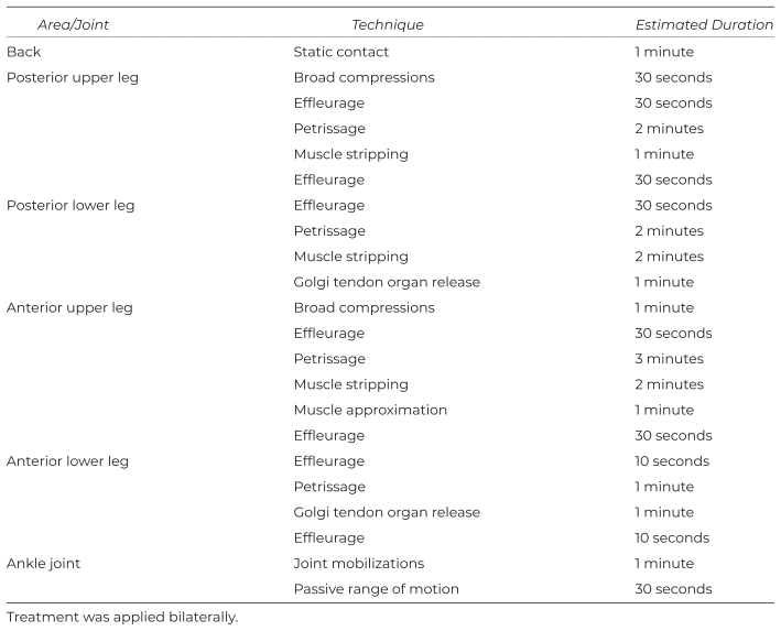
The following techniques were used with consideration of the patient’s goals: static contact, broad compressions, effleurage, petrissage, muscle stripping, Golgi tendon organ release, muscle approximation, joint mobilizations, and passive ROM. Vigorous or painful techniques that may stimulate sympathetic nervous system firing were contraindicated.(27) Static contact was used at the beginning of each treatment to introduce the patient to the therapist’s touch and enhance parasympathetic nervous system activity.(28,29) Broad compressions were performed at a slow rate to assess the level of resting muscle tension and decrease neuromuscular tone.(28) Effleurage was used to spread lubricant, promote lymphatic and venous return, and induce a sedative effect.(28,30,31) Petrissage was used to increase perfusion, decrease resting muscle tension, and decrease motoneuron excitability during application.( 28,32,33) Muscle stripping was used to decrease trigger point activity and restore length to the muscle belly.(28) Golgi tendon organ release and muscle approximation were used to reduce muscle tone and spasticity.(27) Joint mobilizations, consisting of distraction and posterior gliding of the talus at the talocrural joint, and passive dorsiflexion, plantarflexion, supination, and pronation, were used to increase ROM and promote joint nutrition.(27) The treatment plan was first performed on the left side of the patient’s body and then the right; techniques were performed in the order outlined in Tables 1 and 2. The therapist began with general techniques to prepare the tissues for more specific work, and ended with general techniques to promote venous and lymphatic return. The duration of each technique was determined at the discretion of the therapist.
Table 1 Treatment Plan for Weeks 1 and 2
Table 2 Treatment Plan for Weeks 3–6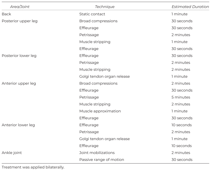
After the second week of treatment, the patient could undress and get on the table quicker than in previous weeks, which allowed for an increase in hands-on time with the patient (see Table 2).
Recommended homecare consisted of a warm bath one to two times per week, and the application of a hot pack to the calves once daily for 10 minutes. Heat has been shown to have a positive effect on spasticity.(34,35)
Verbal and written consent was obtained before the initial assessment. Details discussed included an introduction of the therapist and his credentials, confidentiality, and the risks and benefits of assessment and treatment. The patient was informed that she could decline to participate and could ask questions at any time. During the initial assessment, the therapist and the patient reviewed the clinical findings and established goals of treatment. Verbal consent was obtained at the beginning of each treatment session. The therapist informed the patient that she could alter or stop treatment at any time and explained the treatment to follow, including the patient’s position, areas of the body to be treated, techniques, and rationale for the techniques. Consent was obtained for possible publication of this case report. The patient’s goals and needs were considered when obtaining consent.
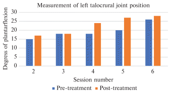
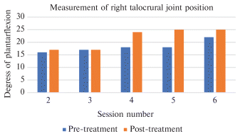
Plantarflexion at rest increased after each treatment and overall from the second to final sessions (see Figures 1 and 2). Left ankle plantarflexion was slightly greater than the right by the end of the treatment. At the final session, slight active dorsiflexion and plantarflexion were possible bilaterally.
|
| ||
|
Figure 1 Measurements of the left talocrural joint angle, taken with the patient seated at the end of the table with her lower leg dependent and foot unsupported. Active range of motion was not possible. Measurements were taken pre- and post-treatment with a universal goniometer. The average of three readings was recorded. No readings were taken during session one. | ||
|
| ||
|
Figure 2 Measurements of the right talocrural joint angle, taken with the patient seated at the end of the table with her lower leg dependent and foot unsupported. Active range of motion was not possible. Measurements were taken pre- and post-treatment with a universal goniometer. The average of three readings was recorded. No readings were taken during session one. | ||
The spasticity of the patient’s knee extensors, measured with the MAS, did not reveal a clinically significant change between the third and last sessions. The MAS score remained a 3 bilaterally throughout all testing. Although the MAS scores remained unchanged, the therapist detected a reduction in spasticity on testing during sessions five and six; the knee went further into flexion before meeting resistance. After each treatment, the therapist could passively move the patient’s ankle back to the pre-treatment start position and slight increases in joint play were noted. The patient expressed that this was the most mobility she could recall having in her ankles. After the final session, the patient demonstrated slight active dorsiflexion and plantarflexion bilaterally.
The patient stated that she experienced greater ease donning her socks and shoes post-treatment and week-to-week. She remarked, “I don’t need to pull on my leg as hard to bend my knee when I put my shoes and socks on, and when I let go of my leg, it doesn’t want to spring back as strong as it did before.” She attended all sessions and reported full compliance with the recommended homecare.
The goal for this case study was to determine the effectiveness of MT in increasing ankle mobility and decreasing spasticity in an adult with spastic CP. This study yielded promising results, particularly given the chronicity of the patient’s condition.
The neuromuscular techniques applied may have caused a reduction in neuromuscular tone due to the mechanical stress placed on proprioceptors, resulting in improved joint mobility and decreased spasticity.(28,33) Joint mobilizations may have contributed to increased mobility as mobilizations stimulate joint proprioceptors, decrease the tone of muscles crossing the joint, and reduce adhesions.(27) Golgi tendon organ release and muscle approximation have been shown to reduce muscle tone and spasm,(27) which could be a factor in the improvement of the patient’s ankle mobility and reduction in knee extensor spasticity.
Left ankle plantarflexion was slightly more than the right. As treatment was the same bilaterally and the difference was minimal, the therapist did not deem the difference to be significant.
No clinically significant changes were detected in the score for spasticity of the knee extensors, which differs from the findings in other studies. In one study examining the effect of MT on spasticity in 20 children with CP, MT was shown to decrease MAS ratings.(13) In another study containing 75 participants, ages 2–10, MAS ratings showed a significant reduction at 6 and 12 weeks.(2) Both studies were conducted over 12 weeks, a considerably longer duration than this study. Additionally, both studies only involved children, which may illustrate the importance of early intervention when considering MT as a treatment for spastic CP. The absence of a clinically significant change in spasticity in this study could be due to the chronicity of the patient’s condition and the limited number of treatments provided. Although there was no change in the MAS score, a reduction in spasticity was detected. The ability to flex the patient’s knee further before meeting resistance during the administration of the MAS and the patient’s reported improvement in function, while not considered clinically significant, should not be overlooked.
There are several limitations in this study. Various massage techniques were used, making it difficult to determine which techniques were effective. Future studies could explore the effectiveness of specific techniques. One person was examined during this study; however, it would be beneficial to conduct future studies with larger sample sizes to obtain more comprehensive data on the effectiveness of MT in adults with CP. No follow-up session occurred; future studies could include a follow-up session 1–2 months after treatment to evaluate sustainability of results. Spasticity of the muscles crossing the ankle joint could not be evaluated using the MAS due to insufficient ROM, making it difficult to determine if ankle mobility improved due to a reduction in spasticity, an increase in joint health, or a combination of both.
Despite limitations, this study demonstrated that MT could have a positive effect on increasing ankle mobility and decreasing spasticity in an adult with spastic CP. It is notable that the patient gained the ability to actively dorsiflex and plantarflex her ankles after decades of limitation, which suggests that MT can positively affect adults with spastic CP regardless of the chronicity of the condition. When considering the duration of this study and the long-standing nature of the patient’s condition, it is remarkable that these results were achieved. It is feasible that longer MT treatments, given over a broader timespan, could have an even greater positive impact on function and mobility in adults with CP. Researchers should be encouraged to conduct further studies on adults with spastic CP; results from future studies will help massage therapists formulate evidence-based treatment plans.
The author would like to thank the staff and faculty of the Massage Therapy Diploma Program at MacEwan University for their help and support throughout the program and this case report. A special thank you to Lois Wihlidal for her ongoing encouragement and guidance.
No sources of funding were used in this study.
The author declares there are no conflicts of interest.
1. CP-NET. About cerebral palsy. http://cpnet.canchild.ca/en/about-cp/about-cerebral-palsy. Accessed January 16, 2023.
2. Mahmood Q, Habibullah S, Babur MN. Potential effects of traditional massage on spasticity and gross motor function in children with spastic cerebral palsy: a randomized controlled trial. Pak J Med Sci. 2019;35(5):1210–1215. https://doi.org/10.12669/pjms.35.5.478
Crossref PubMed PMC
3. Hallman-Cooper JL, Rocha Cabrero F. Cerebral palsy. In: StatPearls. StatPearls Publishing; October 10, 2022. https://www.ncbi.nlm.nih.gov/books/NBK538147/. Accessed February 4, 2023.
4. O’Shea TM. Diagnosis, treatment, and prevention of cerebral palsy. Clin Obstet Gynecol. 2008;51(4):816–828. https://doi.org/10.1097/GRF.0b013e3181870ba7
Crossref
5. Krigger KW. Cerebral palsy: an overview. Am Fam Physician. 2006;73(1):91–100.
PubMed
6. Murphy N, Such-Neibar T. Cerebral palsy diagnosis and management: the state of the art. Curr Probl Pediatr Adolesc Health Care. 2003;33(5):146–169. https://doi.org/10.1016/S1538-5442(03)00002-6
PubMed
7. Gulati S, Sondhi V. Cerebral palsy: an overview. Indian J Pediatr. 2018;85(11):1006–1016. https://doi.org/10.1007/s12098-017-2475-1
Crossref
8. Patel DR, Neelakantan M, Pandher K, Merrick J. Cerebral palsy in children: a clinical overview. Transl Pediatr. 2020;9(Suppl 1):S125–S135. https://doi.org/10.21037/tp.2020.01.01
Crossref PubMed PMC
9. Goldstein M. The treatment of cerebral palsy: what we know, what we don’t know. J Pediatr. 2004;145(Suppl 2):S42–S46. https://doi.org/10.1016/j.jpeds.2004.05.022
Crossref PubMed
10. Kuo CC, Huang HP, Wang TM, Hong SW, Hung LW, Kuo KN, et al. Tendon release reduced joint stiffness with unaltered leg stiffness during gait in spastic diplegic cerebral palsy. PLoS One. 2021;16(1):e0245616. https://doi.org/10.1371/journal.pone.0245616
Crossref PubMed PMC
11. Liptak GS. Complementary and alternative therapies for cerebral palsy. Ment Retard Dev Disabil Res Rev. 2005;11(2):156–163. https://doi.org/10.1002/mrdd.20066
Crossref PubMed
12. Hurvitz EA, Leonard C, Ayyangar R, Nelson VS. Complementary and alternative medicine use in families of children with cerebral palsy. Dev Med Child Neurol. 2003;45(6):364–370. https://doi.org/10.1017/S0012162203000707
Crossref PubMed
13. Hernandez-Reif M, Field T, Largie S, Diego M, Manigat N, Seoanes J, et al. Cerebral palsy symptoms in children decreased following massage therapy. Early Child Dev Care. 2005;175(5):445–456. https://doi.org/10.1080/0300443042000230546
Crossref
14. Majnemer A, Shikako-Thomas K, Shevell MI, Poulin C, Lach L, Schmitz N, et al. Pursuit of complementary and alternative medicine treatments in adolescents with cerebral palsy. J Child Neurol. 2013;28(11):1443–1447. https://doi.org/10.1177/0883073813488942
Crossref PubMed
15. Glew GM, Fan MY, Hagland S, Bjornson K, Beider S, McLaughlin JF. Survey of the use of massage for children with cerebral palsy. Int J Ther Massage Bodywork. 2010;3(4):10–15. https://doi.org/10.3822/ijtmb.v3i4.47
PubMed PMC
16. Wihlidal L. MTST 155: Assessment for Massage Therapists II. Edmonton: MacEwan University; 2022:55–65.
17. Gajdosik RL, Bohannon RW. Clinical measurement of range of motion. Review of goniometry emphasizing reliability and validity. Phys Ther. 1987;67(12):1867–1872. https://doi.org/10.1093/ptj/67.12.1867
Crossref PubMed
18. Swann E, Harrelson GL. Measurement in rehabilitation. In: Andrews JR, Harrelson GL, Wilk KE, eds. Physical Rehabilitation of the Injured Athlete. Elsevier; 2012:67–73. https://doi.org/10.1016/B978-1-4377-2411-0.00005-8
Crossref
19. Ekstrand J, Wiktorsson M, Oberg B, Gillquist J. Lower extremity goniometric measurements: a study to determine their reliability. Arch Phys Med Rehabil. 1982;63(4):171–175.
PubMed
20. Meseguer-Henarejos AB, Sánchez-Meca J, López-Pina JA, Carles-Hernández R. Inter- and intra-rater reliability of the Modified Ashworth Scale: a systematic review and meta-analysis. Eur J Phys Rehabil Med. 2018;54(4):576–590. https://doi.org/10.23736/S1973-9087.17.04796-7
Crossref
21. Morris S. Ashworth and Tardieu Scales: their clinical relevance for measuring spasticity in adult and paediatric neurological populations. Phys Ther Rev. 2002;7(1):53–62. https://doi.org/10.1179/108331902125001770
Crossref
22. Pandyan AD, Johnson GR, Price CIM, Curless RH, Barnes MP, Rodgers H. A review of the properties and limitations of the Ashworth and Modified Ashworth Scales as measures of spasticity. Clin Rehabil. 1999;13(5):373–383. https://doi.org/10.1191/026921599677595404
Crossref PubMed
23. Fosang AL, Galea MP, McCoy AT, Reddihough DS, Story I. Measures of muscle and joint performance in the lower limb of children with cerebral palsy. Dev Med Child Neurol. 2003;45(10):664–670. https://doi.org/10.1017/S0012162203001245
Crossref PubMed
24. Bohannon RW, Smith MB. Interrater reliability of a Modified Ashworth Scale of muscle spasticity. Phys Ther. 1987;67(2):206–207. https://doi.org/10.1093/ptj/67.2.206
Crossref PubMed
25. Validity of Outcome Measures. Canadian Agency for Drugs and Technologies in Health; 2018. https://www.ncbi.nlm.nih.gov/books/NBK540254. Accessed February 28, 2023.
26. Ghotbi N, Ansari NN, Naghdi S, Hasson S, Jamshidpour B, Amiri S. Inter-rater reliability of the Modified Modified Ashworth Scale in assessing lower limb muscle spasticity. Brain Inj. 2009;23(10):815–819. https://doi.org/10.1080/02699050903200548
Crossref PubMed
27. Rattray FS, Ludwig L. Clinical Massage Therapy: Understanding, Assessing and Treating Over 70 Conditions. Talus Incorporated; 2000.
28. Andrade CK. Outcome-Based Massage: Putting Evidence into Practice. 3rd ed. Philadelphia: Wolters Kluwer Lippincott Williams & Wilkins; 2014.
29. Post-White J, Kinney ME, Savik K, Gau JB, Wilcox C, Lerner I. Therapeutic massage and healing touch improve symptoms in cancer. Integr Cancer Ther. 2003;2(4):332–344. https://doi.org/10.1177/1534735403259064
Crossref
30. Monteiro Rodrigues L, Rocha C, Ferreira HT, Silva HN. Lower limb massage in humans increases local perfusion and impacts systemic hemodynamics. J Appl Physiol. 2020;128(5):1217–1226. https://doi.org/10.1152/japplphysiol.00437.2019
Crossref PubMed
31. Harris M, Richards KC. The physiological and psychological effects of slow-stroke back massage and hand massage on relaxation in older people. J Clin Nurs. 2010;19(7–8):917–926. https://doi.org/10.1111/j.1365-2702.2009.03165.x
Crossref PubMed
32. Morelli M, Chapman C, Sullivan SJ. Do cutaneous receptors contribute to the changes in the amplitude of the H-reflex during massage? Electromyogr Clin Neurophysiol. 1999;39(7):441–447.
PubMed
33. Goldberg J, Seaborne DE, Sullivan SJ, Leduc BE. The effect of therapeutic massage on H-reflex amplitude in persons with a spinal cord injury. Phys Ther. 1994;74(8):728–737. https://doi.org/10.1093/ptj/74.8.728
Crossref PubMed
34. Denton A, Bunn L, Hough A, Bugmann G, Marsden J. Superficial warming and cooling of the leg affects walking speed and neuromuscular impairments in people with spastic paraparesis. Ann Phys Rehabil Med. 2016;59(5–6):326–332. https://doi.org/10.1016/j.rehab.2016.04.006
Crossref PubMed
35. Matsumoto S, Kawahira K, Etoh S, Ikeda S, Tanaka N. Short-term effects of thermotherapy for spasticity on tibial nerve F-waves in post-stroke patients. Int J Biometeorol. 2006;50(4):243–250. https://doi.org/10.1007/s00484-005-0009-4
Crossref
Table S1 Modified Ashworth Scale Scoring (24)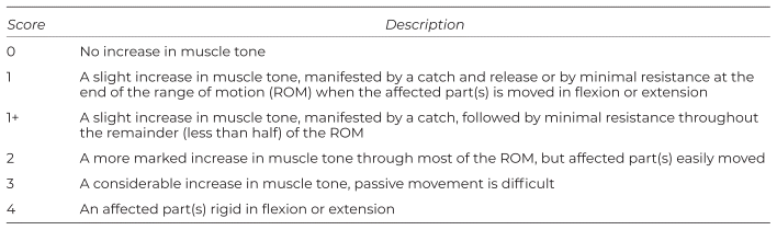
Corresponding author: Spencer Pon, MacEwan University Massage Therapy, MacEwan University, 9-302F, 10700 - 104 Avenue NW, Edmonton, AB T5J 4S2, Canada, E-mail: spencerponrmt@gmail.com, Tel: +(780) 499-1975
COPYRIGHT
Published under the CreativeCommons Attribution-NonCommercial-NoDerivs 3.0 License.
International Journal of Therapeutic Massage and Bodywork, Volume 18, Number 1, March 2025